Clinical image
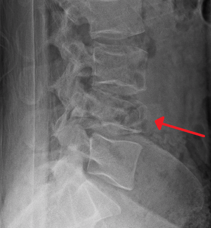
Lateral radiograph showing an L4 burst fracture. Source: Wikimedia Commons — James Heilman, MD; CC BY-SA 4.0. Image binary is embedded locally in this package; original source and licence are retained.

Key points
Talk
- I can see AP and Lateral radiograph of lumbar spine. AP X-ray show widening of inter pedicular distance at L1 and Lateral shows compression of the L1 vertebra.
- I will approach the patient along ATLS protocol and BOAST guideline for SCI and Spine clearance.
- Once initial assessment and management is completed I will classify the Fracture based on TLICS system by clinical examination and multi directional imaging including CT and MRI
TLICS is a classification system in which integrity of posterior ligamentous structures and neurological status are taken in to consideration, along with the fracture morphology
This system aids surgical decision making 5 or more score treat operatively