Clinical image
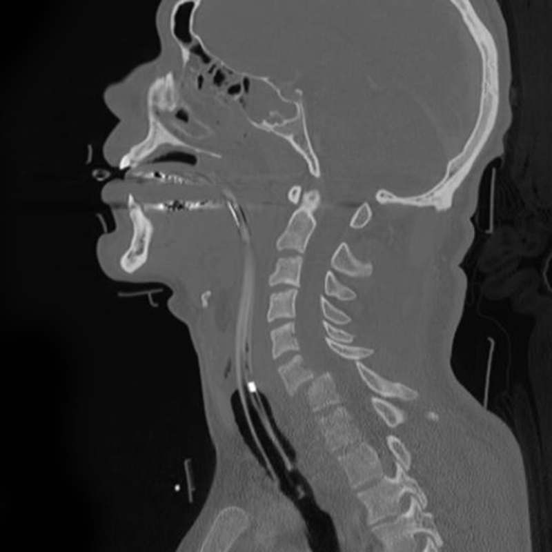
Sagittal CT reconstruction of a C6–C7 fracture-dislocation. Source: Wikimedia Commons — Frank Gaillard; CC BY-SA 3.0. Image binary is embedded locally in this package; original source and licence are retained.

Talk
- I will approach the patient based on ATLS protocol and BOAST guideline for SCI. It involves
- Initial assessment
- Initial management
- Definitive management
- Post management followup
Initial Assessment
- Immobilise the spine until investigations are over and c spine is cleared
- ASIA chart recorded
- Identify spinal shock
- Differentiate neurogenic from hypovolemic shock
- Imaging based on BOAST 2 guideline
Initial management
- Protect
- Spine
- Position flat
- GI
- Genitourinary
- Skin
- DVT
- Physio
Definitive management
- Occypital condyle #
- I:Compression
- II:direct blow
- III: Rotary
- I and II collar
- III: O-C2 fusion
Treatment
- Stable: Rigid collar
- Unstable: Combine lateral mass distance 7 or more mms
- Traction and Halo
- O-C2 Fusion
AAI
Adult
- Degnerative: RA, Downs
- Trauma
Paediatric
- Degenerative: JRA, Downs
- Trauma
- Infection
AADI >5MM
SAC <9MM
MRI to be done
Stable, children: non surgical
Unstable: C1-C2 fusion
Odontoid Fracture
Transverse most important
Odontoid
- Bimodal distribution
- D/D os odentoidum (CT)
- 1,3: non surgical (rigid collar, Halo)
- 2: high risk of nonunion
- Halo
- Surgery
- anterior stabilisation (AS-PI fracture pattern)
- posterior stabilisation (Operating close to Vertebral artery), ct angeo
Hangman
Traumatic S/L of C2 over C1
- I: <3mm displacement Rigid collar
- II: > 3mm displacement Halo after CR
- IIA: angulation with out displacement Halo after CR/Never traction
- III: Facet joint dislocation closed/open reduction and stabilise
Subaxial
- Flexion compression
- Flexion distraction
- Extension compression
- Extension distraction
- Vertical compression
- Lateral rotation
Flexion distraction
Facet dislocation and Fracture
- Request an MRI
Facet dislocation and Fracture](Cervical trauma_assets/slide-15-01.jpg)
U/L
- <25%
- Mono radiculopathy
- Superior facet fracture
B/L
50%
- Spinal cord symptoms
- B/L radiculopathy
Closed reduction
- I will do MRI in all cases
- Discuss with National spine injury unit
- In theatre
- adequate anaesthesia
- C arm cross table lateral
- Spine unit back up
- Garden well tong, weight is based on patients body wt
- Stop if neurology worsens, do repeat MRI
Refer to Spine unit
- Anterior disc in MRI
- Failed C/R
Obtunded
Anterior approach
- Mandible: c2
- Hyoid: c3
- Thyroid: C4-5
- Cricoid: c6
Position
- Supine
- Sandbag under shoulder
- Face turned away
- Longitudinal skin incision along anterior border of SM
- Platisma split longitudinally
- SM laterally, Sternohyoid and Sternothyroid muscle retracted medially
- retract carotid sheet laterally by blunt dissection
- Ligate Sup and Inf Thyroid arteries
- Split pre vertebral fascia and sub per elevate along with longs coli
Lt or Rt
- Left recurrent L N is arising from vagus as it cross arch of aorta
- Rt RLN arise from vagus as it cross 1st part of subclavian
- Rt RLN can be aberrant rarely
- thoracic duct is on left