Clinical image
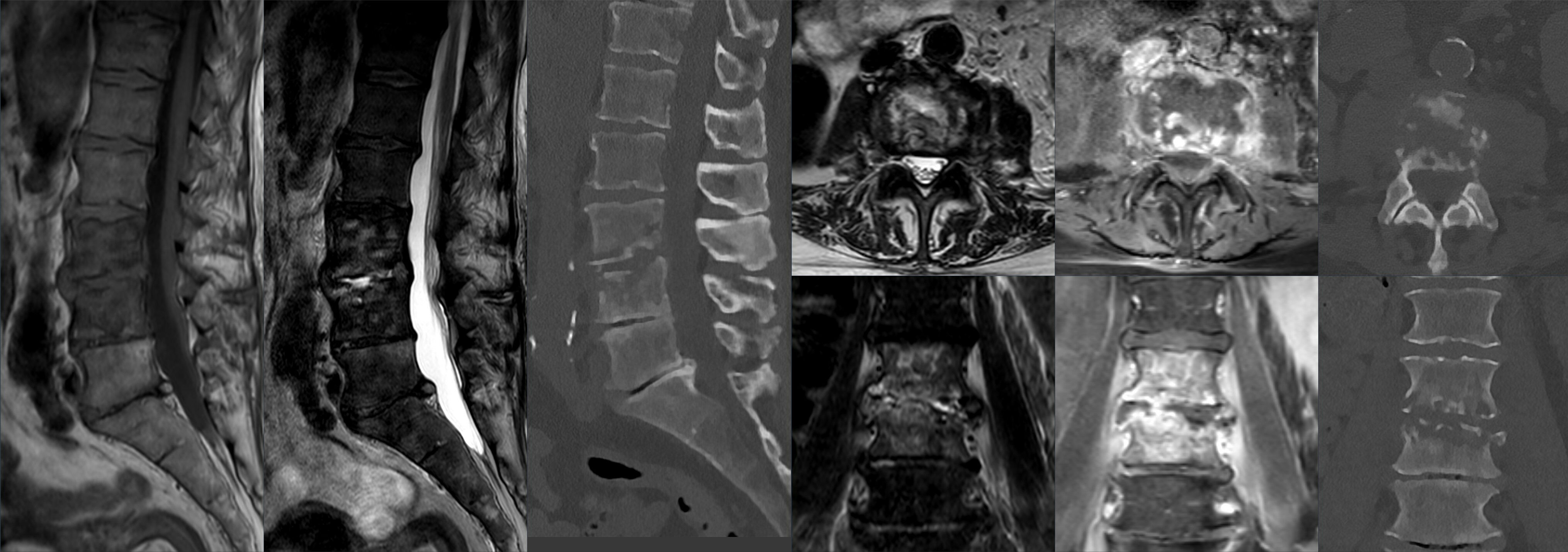
MRI and CT appearances of lumbar spondylodiscitis. Source: Wikimedia Commons — Hellerhoff; CC BY-SA 4.0. Image binary is embedded locally in this package; original source and licence are retained.

Talk
- T2 weighted image A shows high signal from C6 and C7 vertebral bodies with reduced disk height. There is a region of high T2 signal anterior to the vertebrae.
- T1 weighted probable Gad enhanced MRI gives the same picture with enhancement
- This is highly suggestive of epidural abscess.
- I will take a detailed history looking for the predisposing factors, comorbiditis and addictions. I will do a complete spine examine and assess for neurological status .
- I will start immediate empirical antibiotic and prepare patient for emergency debridement and culture and biopsy
Definitions
- Infection of IVD : spondylodiskitis
- Infection of endplate or body: V OM/spondylitis
- Infection of canal: Epidural abscess
Children vs adults
- NO bone infarct in children
- Extensive intraoseous anasthemosis
- Disk is vascular
- Bone infarct, erosions and osteophytosis: Adult
- Disk avascular
- Less anasthemosis
Key points
Non surgical
- Targeted antibiotic
- Immobilisation to
- relieve pain and
- help preventing instability and deformity
- Rest
Indication for surgery
- Neurology
- Instability
- Sepsis
- Epidural abscess
Surgical Procedure
- Anterior debridement
- Reconstruction with bone construct
- Posterior decompression
- Instrumented stabilisation