Standard radiographs
- Weight-bearing AP knee
- Lateral knee
- Skyline / Merchant view
- Full-length standing AP radiograph when overall mechanical alignment is being assessed
Alignment
Assess the overall coronal alignment first. On the full-length AP radiograph, the mechanical axis is assessed from the centre of the femoral head through the knee to the centre of the ankle. Component position should then be assessed independently.
Femoral component
- Coronal alignment on AP view
- Flexion or extension on lateral view
- Anterior femoral notching
- Overhang or undercoverage
Tibial component
- Varus or valgus position on AP view
- Posterior slope on lateral view
- Tibial tray coverage and overhang
- Subsidence or migration
Joint line
Joint-line restoration should be assessed on the AP radiograph. Elevation of the joint line may alter knee kinematics, flexion-gap behaviour and patellofemoral mechanics.
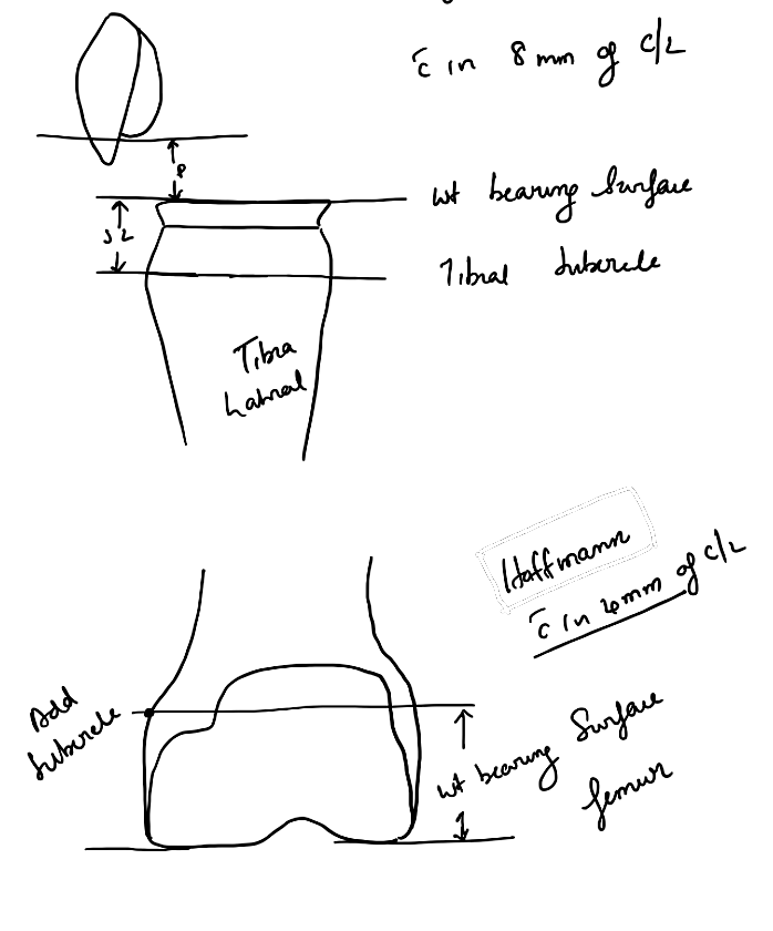
The joint line may be estimated in relation to reproducible bony landmarks such as the fibular head, tibial tubercle and adductor tubercle. Comparison with the contralateral knee or pre-operative films is useful when available.
Radiolucent lines and loosening
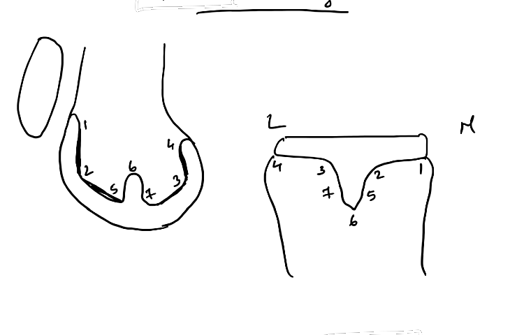
Assess the bone–cement or bone–implant interface systematically on AP and lateral views.
A narrow, non-progressive radiolucent line can occur around a stable implant. Features that support loosening are:
- Progressive radiolucent lines
- Increasing width of the interface
- Component migration or change in position
- Subsidence
- Progressive osteolysis
Radiolucencies should be recorded by zone rather than described vaguely.
Patellofemoral assessment
The skyline view is used to assess:
- Patellar tilt
- Lateral subluxation
- Patellar component position
- Symmetry of patellar resection
- Patellofemoral overstuffing
Maltracking may result from component malposition, particularly internal rotation of the femoral or tibial component, an increased Q angle, or abnormal patellar component position.
Component rotation
Plain radiographs are limited for assessing rotational alignment. CT is used when component malrotation is suspected, particularly in a painful TKA with stiffness or patellar maltracking.
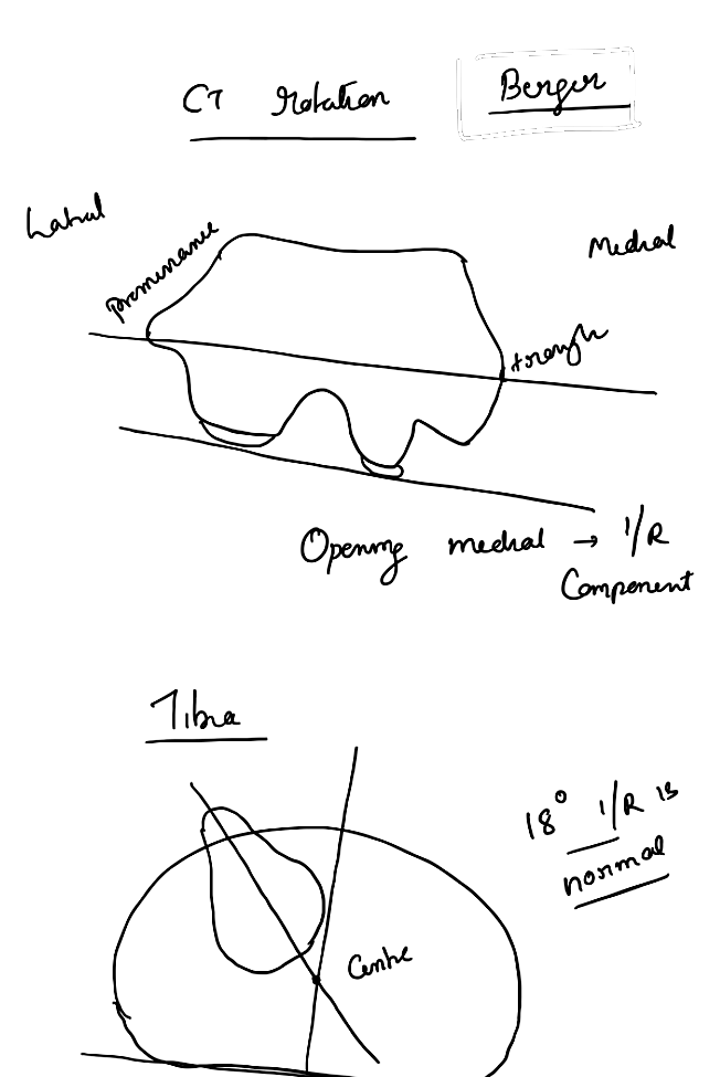
Femoral rotation is assessed in relation to the transepicondylar axis and the posterior condylar axis. Tibial rotation is assessed using the tibial component axis in relation to the tibial tubercle and other established tibial reference axes.
Lateral view
Assess:
- Femoral component flexion or extension
- Anterior femoral notching
- Posterior femoral condylar offset
- Tibial slope
- Component subsidence or migration
- Patellar height
- Posterior osteolysis
Features to look for in a painful TKA
- Aseptic loosening
- Periprosthetic joint infection
- Instability
- Component malposition or malrotation
- Polyethylene wear and osteolysis
- Patellar maltracking
- Periprosthetic fracture
- Extensor mechanism abnormality
References
- Orthobullets. TKA Axial Alignment.
- Orthobullets. TKA Patellofemoral Alignment.
- Orthobullets. Adult Knee Radiographic Evaluation.
- Orthobullets. TKA Aseptic Loosening.