Clinical image
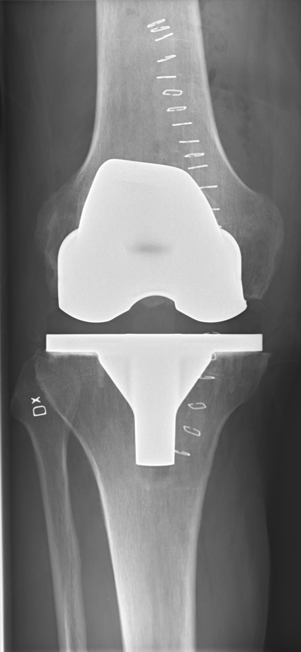
Postoperative AP radiograph of a total knee prosthesis. Source: Wikimedia Commons — Mikael Häggström, M.D.; CC0 1.0. Image binary is embedded locally in this package; original source and licence are retained.

Reported success rate
- Overall 14 year Revision rate is 4.47%
- CR 4.0 %
- PS 5.4%
- UKA 9%
- PFJ 25%
Indication
- Pain not relieved by conservative measures, affecting ADL
C/I
- Infection
- Neurological disease
- Vascular disease
- Deficient quadriceps
Surgical goal
- Pain relief
- Functional improvement
Technical goals
- Achieve wt bearing axis through the centre of knee
- Joint line Perpendicular to Wt bearing axis
- Soft tissue balancing
- Restore the Q angle
- Joint level
Valgus cut angle
- Angle between mechanical and anatomical axis
- Mechanical axis is at 1.2 degree varus.
- So 60% weight passes through medial compartment
Femoral roll back
- Posterior shift in the FT contact point in the sagittal plain as the knee flexes
Constraint ladder
- When constrain increases, conformity and there by aseptic loosening increases.
- PCL retaining
- PCL retaining with mobile bearing
- PS
- Constrained condylar non hinged (unlinked)
- Constrained condylar hinged (Linked)
Key points
| PCL retain | PS |
|---|---|
| Least constrained | More constrained |
| Less loosening | Less PE Delamination |
| Preserve bone stock | Femoral bone loss |
| Preserve proprioception | AP stability due to cam |
| S/C # easy to manage | Cam jump |
| Poor roll back | Better roll back |
| More PE delamination | More Loosening |
| PCL rupture & Flexion instability | Patella clunk sy |
Medio lateral Balancing
| Medial release | Lateral release |
|---|---|
| Osteophyte | Osteophyte |
| Deep MCL | PF ligament |
| SM | PL capsule |
| Superficial MCL | LCL (Both F and E tight) |
| Pes anserina | ITB, Popliteus (Ext tight) |
| Biceps and Gastro (Flex only) | |
| PCL | PCL |
Flexion extension balancing
| Column 1 | Column 2 | Column 3 | Column 4 |
|---|---|---|---|
| Flexion Loose | Flexion OK | Flexion tight | |
| Extension loose | Thick insert | Augment femur | Down size femur and thick insert |
| Extension OK | Oversize Femur | Perfect | Down size femur |
| Extension Tight | Oversize femur and thin insert | Resect femur | Cut tibia |
Tibial cut
- 3-5 degree posterior slope
- Perpendicular to mechanical axis
- Use EM jig
Distal femoral cut
- Perpendicular to mechanical axis
- Achieved by 5-7 degree of VC angle
- Use IM jig
AP femoral cut
- Tibial is in 3 degree varus naturally
- ER of femoral cutting block to 3 degree
- Achieved by
- keeping jig parallel to epicondylar axis
- Posterior referencing
- Perpendicular to whitesides line
Patello femoral Maltracking
- Prevented by
- ER Femoral component
- ER Tibial component
- Lateralise FC
- Lateralise TC
- Medialise Patellar component
Patella resurfacing
- RCT, Roberts et al: Improved result with electrocautery than none
- RCT, Barrack et al: Vast majority of patients with remaining patellar articular cartilage do well regardless of resurfacing
- In view of above two studies, I will selectively resurface. In grade 3-4 OA of patella, I would resurface. For rest of patients I will do circumferential electrocautery.
Managing Patella baja
- Use small patella dome
- Lower joint line
Key points
Talking the talk
- AP radiograph of both knees showing bilateral lateral compartment degenerative changes which is more in the right side. There are valgus with loss of joint space in the lateral compartment, with sclerosis and osteophyte formation.. These findings are consistent with OA knees
- I would take a detailed history trying to establish the cause and success of conservative measures.
- I will examine her gait, will do a full knee examination to determine the severity of deformity. I will assess the integrity of collateral ligaments and correct-ability of deformity to plan for the degree of constrain needed. I will also examine her hips and ankles for any deformity or stiffness.
- I will also obtain a lateral view and skyline view. A long leg view would help me to determine the Mechanical axis and degree of valgus. complete BC, CRP and ESR is done to rule out infection if any surgical intervention is planned
Causes
- Inflammatory joint disease
- Female
- Post traumatic
- osteonecrosis
- HTO
Valgus Knee
- TF angle > 10 degree
- Associate with bony and soft tissue abnormalities
- Lateral subluxation of patella due to Lateral capsule and lig contracture: Large insert and chance of perineal nerve palsy
- Medial laxity: Constrained knee may be needed
- PCL elongated and dysfunctional: PS Knee
- Deficient Lateral condyle posteriorly and Distal femoral IR rotation up to 10 degree: Posterior referencing C/I
Principles
- Medial parapatellar incision
- Distal femoral cut using predetermined Valgus cut angle
- Proximal tibial cut with 3 degree posterior slope
- Mediolateral balancing
- AP femoral cut parallel to epicondylar axis or perpendicular to whiteside line