Clinical image
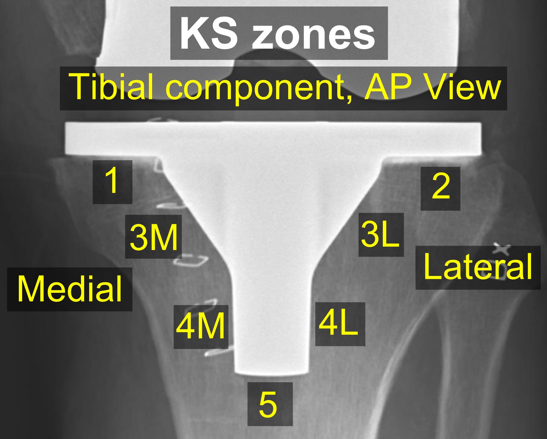
AP radiograph of a knee prosthesis showing Knee Society radiographic zones. Source: Wikimedia Commons — Mikael Häggström, M.D.; CC0 1.0. Image binary is embedded locally in this package; original source and licence are retained.

Key points
Talking the talk
- AP and Lateral radiograph showing PS TKA. Both components appear well fixed. My primary concern is an area of subchondral lucency under the medial tibial component. I will suspect infection unless otherwise proved but will rule out other causes of painful TKA
- Causes of painful tka are Intrinsic, extrinsic and Infection
- I will take a detailed history and examination stressing on
- the primary indication,
- post operative course including leaking wounds,
- pattern of pain,
- Health of scar and
- the skin condition and
- instability.
Causes
- Infection
- Intrinsic causes: AIMS and 3 Fractures
- Aseptic loosening
- Instability
- Malalignment
- Stiffness, soft tissue impingement
- Patellar problems
- Extensor mechanism rupture
- Periprosthetic fracture
- Implant breakage
- Extrinsic causes
- Hip pathology
- Neurologic
- Spine Pathology
- CRPS
- Vascular
- Unexplained 1%
Aseptic loosening
- Decreased joint space
- Radiolucency
Instability
- ML
- AP
- F/E
Malalignment
- Sagital
- coronal
- Rotational
CFA: Coronal femoral component angle, CTA: coronal TC angle
Soft tissue impingement
- Component overhang
- Patellar clunk
- Popliteal tendon impingement
- Fabellar impingement
Overhang
Clung syndrome
History
- Preop
- Indications
- Pain
- Expectations
- Depression and anxiety
- Wound problems
Symptoms
- Pain
- Swelling
- Stiffness
- Instability
Pain
- Unchanged (never felt right)
- Sharp catch (impingement)
- Painful to touch (CRPS)
- Start up pain (Aseptic loosening)
- Getting down stairs (PF)
C/E
Gait (Thrust)
Skin: inflammation, sinus, atrophy (CRPS)
Effusion
Hip
Tenderness
PF
Medial: overhang, pes anserina
PM: Popliteus
Cutaneous neuroma
ROM
PF joint
Spin hip and foot and ankle, NV
Inv
- xray: AP, Lateral, skyline
- Alignment
- Loosening
- Wear of PE
- Overhanging
- Joint line
- Fractures
CT
- Malrotation
Bone scan
- Good negative
Lab
- FBC
- ESR >30 (3 mo post op)
- CRP> 10(3 weeks)
- IL 6 > 10pico gram
Syovial fluid aspiration
- All painful knee
- Gram
- WBC >3000 a/c, 10000 in c/c
- 2 weeks no Ab
- Leukocyte esterases
- Alfa defincin
PJI
- Approach
- Difficult problem
- Local bone infection unit
- MDT
- 1-2%
Key points
AAOS GUIDELINE for KNEE
- Test ESR (75% Sn, 70 % Sp) , CRP (88 and 74)
- IL 6 (97 and 91)
- Aspirate
- Tissue culture: 3-6 using different instruments from diff areas
- No Ab until after culture
- No Gram stain inta operative
- Nuclear imaging
Classification
- a/c: <3 weeks
- c/c: >3 weeks
Management
Biofilm
- A polysaccaride film ( glycocalyx)
- improves bacterial nutrition
- Reduce access of Ab to bacteria
DAIR: < 3 WEEKS
2 stage revision
- gold std
- 1st stage: debride, spacer, ab for 6 weeks
- 2nd stage:> 5 weeks( CRP and culture neg)
Others
- Single stage
- Long term Ab suppression
- Arthrodesis
- Amputation/disarticulation
2 vs 1 stage
- Jamsen et al: equal functional result with better infection eradication in 2 stage
- Buechel et al: 90% infection eradication with 1 stage