Origin
The ulnar nerve is a terminal branch of the medial cord of the brachial plexus, principally carrying fibres from C8 and T1.
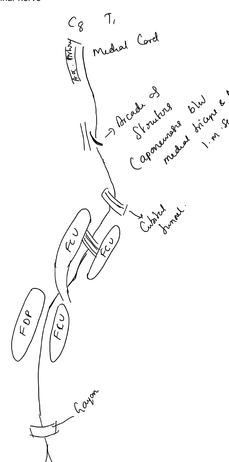
Course
Arm
- Runs medial to the brachial artery proximally
- Pierces the medial intermuscular septum in the distal arm
- Passes posterior to the medial epicondyle
Elbow
The nerve passes through the cubital tunnel behind the medial epicondyle.
Forearm
- Enters between the two heads of flexor carpi ulnaris
- Runs between FCU and flexor digitorum profundus
Wrist and hand
- Passes superficial to the flexor retinaculum through Guyon canal
- Divides into superficial sensory and deep motor branches
Motor supply
Forearm
- Flexor carpi ulnaris
- Ulnar half of flexor digitorum profundus
Hand
- Hypothenar muscles
- Interossei
- Third and fourth lumbricals
- Adductor pollicis
- Deep head of flexor pollicis brevis
Sensory supply
- Little finger
- Ulnar half of ring finger
- Corresponding ulnar aspect of palm and dorsum through palmar and dorsal cutaneous branches
Clinical examination
- Finger abduction and adduction
- Froment sign
- Wartenberg sign
- First dorsal interosseous strength
- Sensation in little finger
Common sites of compression
- Cubital tunnel
- Guyon canal
A lesion at the elbow affects FCU and FDP in addition to intrinsic hand muscles; a lesion at the wrist spares the forearm muscles.
References
- Orthobullets. Ulnar Nerve.