Organisation of skeletal muscle
Skeletal muscle is composed of muscle fibres containing myofibrils. Each myofibril consists of repeating sarcomeres, which are the functional contractile units of skeletal muscle.
Thin filament
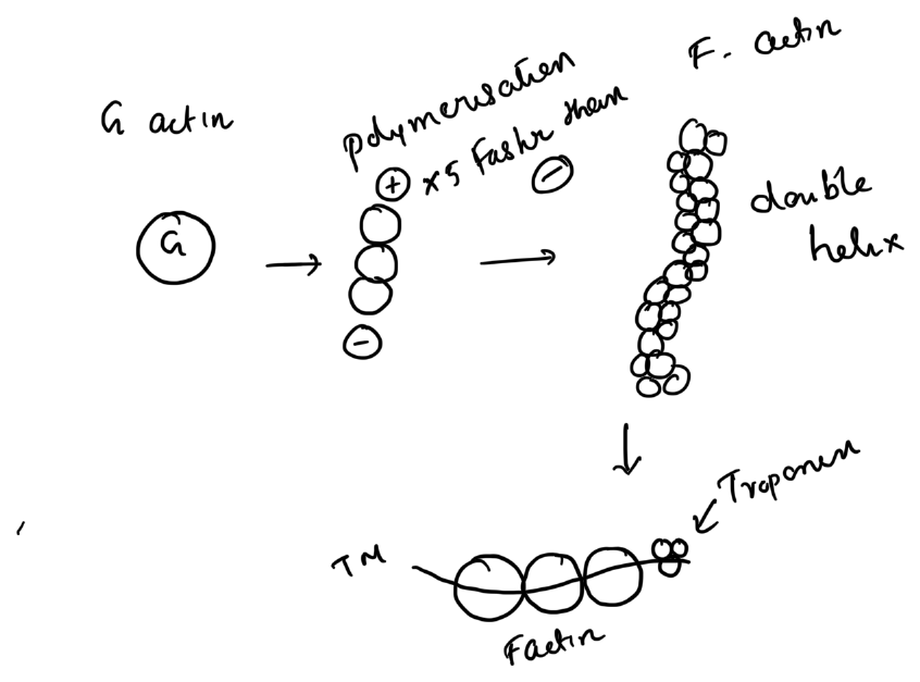
The thin filament contains:
- Actin
- Tropomyosin
- Troponin complex
G-actin monomers polymerise to form F-actin, which is arranged as a double helix. Tropomyosin lies along the actin filament and covers the myosin-binding sites at rest.
The troponin complex consists of:
- Troponin C — binds calcium
- Troponin I — inhibitory component
- Troponin T — binds tropomyosin
Thick filament
The thick filament is composed mainly of myosin. Each myosin molecule has a tail and two globular heads. The heads bind actin and possess ATPase activity.
Sarcomere
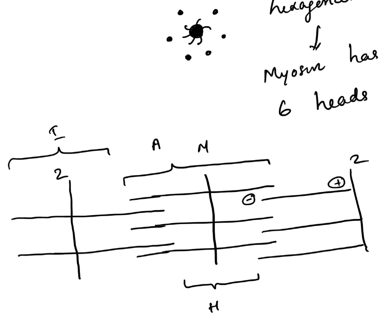
A sarcomere extends from one Z line to the next.
- A band — length of the thick filament; remains constant during contraction
- I band — thin filaments only; shortens during contraction
- H zone — central thick-filament-only region; shortens during contraction
- M line — centre of the sarcomere
Sliding filament mechanism
- An action potential triggers calcium release from the sarcoplasmic reticulum.
- Calcium binds troponin C.
- Tropomyosin moves away from the myosin-binding sites on actin.
- Myosin heads bind actin to form cross-bridges.
- The power stroke pulls the thin filament toward the centre of the sarcomere.
- ATP binding detaches myosin from actin.
- ATP hydrolysis re-cocks the myosin head for another cycle.
During contraction the sarcomere shortens, but the actin and myosin filaments themselves do not shorten.
Length–tension relationship
Maximum active force is generated when actin and myosin overlap optimally. Excessive shortening or excessive lengthening reduces the number of effective cross-bridges and decreases force generation.
Clinical relevance
These principles underlie:
- muscle weakness after immobilisation
- tendon transfer tensioning
- the effect of muscle length on force generation
- contracture and rehabilitation after injury
References
- Orthobullets. Basic Science — muscle and tendon physiology principles.