Clinical image
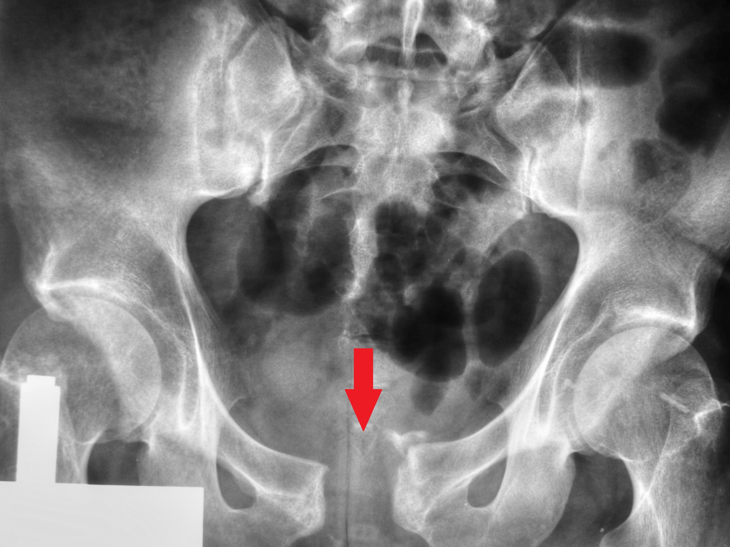
AP pelvic radiograph showing pubic symphysis diastasis. Source: Wikimedia Commons — Nevit Dilmen; CC BY-SA 3.0. Image binary is embedded locally in this package; original source and licence are retained.

Prehospital
- Stop bleeding
- TXA
- Binder
- Hyperfibrinolysis and coagulopathy of a/c trauma
Key points
Talking the talk
- Pelvis X-ray AP view shows an open book APC type III pelvic fracture. My initial concern is the potential
- life threatening nature of the injury because of the increased incidence of vascular injury, abdominal and urogenital injuries associated
- I will follow the ATLS and BOAST 3 guidelines for the initial management.
Principles
- My principles of initial management after securing the airway , breathing and taking AMPLE history are
- To compensate for the blood loss by giving O neg blood in 1:1:1 ratio by activating MTP (based on the MOI, Physiological status of patient and identified injuries)
- To stop the bleeding by homeostatic resuscitation, permissive hypotension and DCS
- Prevent trauma triad of death (Hypothermia, coagulopathy and metabolic acidosis)
- Rule out open pelvic fracture and urologenetal injury
Prevent resuscitation ass coagulopthy
Trauma death triad
Compenstiong Haemorrhage
- MTP: Transfusion of total blood volume in 24 hours
- Early transfusion of warmed blood along with platelets and fresh frozen plasma in a 1:1:1 ratio
- ROTEM and Thromboelastogram: Helps in targeted blood product resuscitation for ongoing blood therapy, based on viscoelastic behaviour of blood clot.
Key points
Permissive hypotension
- SBP maintained at 90mmHg
- Balance between too much blood loss due to high pressure and tissue hypo perfusion due to low pressure
Haemostatic resuscitation
- Primary clot is the best clot and should be maintained for further consolidation of clot
- Pelvic binder applied at the level of GT with internal rotation of both hips and flexion of knees.
- IV Tranexamic acid 1gm given ideally <1hr at least with in 3 hours of injury as bolus followed by infusion over 8 hours (CRASH 2 trial reduce RR of death to .85)
Tranexamic Acid
- Antifibrinolytic
- Completely inhibits the conversion of plasminogen to plasmin thus preventing degradation of fibrin
- Civilian and military studies (CRASH 1 and 2, MATTERs) has demonstrated significant benefit when given with in 3 hours.
DCS
- Identify the source of bleeding
- Bleeding can be in the floor and 4 more: Chest, abdomen, pelvis and thigh.
- External bleeding controlled by direct compression
- FAST can identify free fluid in abdomen
- CT with IV contrast from head to mid thigh and a he’d to toe scanogram is taken based on patient status
- Selective embolisation of arterial bleed
- Visceral bleed: laparotomy or thoracotomy
3 different responses
- Responders: Pelvic binder protocol
- Should not be left >24
- Ex Fix if definitive Sx is delayed (+ reaction if Vertical shear)
- Post binder xray (masking)
- Transient responders and Non responders:
- Surgical packing of pelvis
- Selective embolisation IR
DCO
- CR of dislocations
- Reduce and splint fractures
- Traction
- Ex Fix long bones
- Open fracture : Debride and ex fix
When to do DCO
- Aim is to prevent second hit phenomenon where the condition of patient worsens after a surgery
- Patients are categorised in to Stable, Borderline, Unstable and in extremis after initial resuscitation
- Tests for adequacy of resuscitation and perfusion
- Stable harm-dynamics
- No hypoxemia, hypercapnia
- S.Lactate
- <2mmol/l: ETC
- 2-2.5: observe the trend,
2.5:DCS
- normal coagulation
- normothermia
- normal renal function >1ml/kg/hr
Associated injuries
- Potential injury to bladder and urethra: Follow BOAST 14 guideline
- Potential anorectal injury
Open pelvic fracture
- Urgent assessment by the General surgeon to rule out potential anorectal injury
- Such injury require construction of de functioning stoma.
Urological injury
- Urological trauma is rare and the incidence of severe urethral trauma is 1/million population/year.
- The majority of cases are due to blunt high-energy trauma with associated multi-system injuries and 80% of these cases are associated with pelvic fractures.
- Urological injuries are potentially fatal and can result in severe long-term disability.
Management principles
- Initial
- Theatre
- post operative
Initial
- All high energy injury patients mush have exam of perineum , genitalia and rectum
- Single gentle attempt at catheterisation with 16F soft silicon catheter by an experienced doctor
- Blood stain urine, do retrograde cystogram
- Catheter does not pass/ drains only blood: do not inflate, withdraw and do Retrograde urethrogram
- Inform urologist/paediatric urologist
- Percut, suprapubic catheter by seldinger technique with skin incision in midline 3-4 fingers above symphysis
- Pelvic fracture is treated as open if urine leaks (IV ABx and early surgery)
Retrograde Urethrogram:
- Usually in Resuscitation room.
- X-ray plate under pelvis.
- 20 ml dilute IV contrast medium (10 ml contrast + 10 ml saline).
- Balloon of small Foley catheter into penile meatus and gently inflated.
- Hold catheter in place and inject contrast.
- AP Pelvis x-ray taken. Additional lateral if possible.
Catheter Cystogram:
  - • Usually in Resuscitation room.
- X-ray plate under pelvis.
- 300ml dilute IV contrast medium (150 ml contrast + 150 ml saline).
- Push catheter in a further 2-3 cm so balloon not blocking bladder neck.
- nject contrast down catheter with bladder syringe and clamp catheter.
- AP Pelvis x-ray taken. Additional lateral if possible.
- Evacuate contrast and repeat AP Pelvis x-ray.
Theatre
- Intraperitoneal bladder rupture: emergency laparotomy and direct repair
- Extraperetoneal bladder
- Stable pelvis: catheter only
- unstable: fix fracture and repair bladder
- Identified during pelvic sx: repair
- Urethras injury
- Children and female: supra regional urologist discussion
- Primary repair: with anorectal inj, perennial devolving, bladder neck inj, massive bladder displacement, penetration of anterior urethra
- Male: delayed repair 3 months
Post operative
- Communicate high incidence of urinary and sexual dysfunction
- Linked andrological service
- TRAN and audit
Definitive treatment Pelvis
- Referred to specialist centre with in 24 hours
- Reconstruction with in 72 hours
- Thrombo prophylaxis
- Follow up for Functional recovery
- Written advice on sexual disfunction
Definitive treatment
- Investigations:
- inlet and outlet view
- CT
- Classification: Young and burgess
- A: LC Rami #
- sacral ala #
- post ilium #
- 11 with C/L posterior injury
- B: APC
- APC1: <2.5 cm pubic widening
- APC 11: >2.5 cm (post lig intact)
- APC111: anterior and post big gone. High vascular injury
- C: VS
- A: LC Rami #
Treatment
- Non surgical: Undisplaced #/Patient too ill for surgery
- Operative :
- Plate fixation of Pubic diasthesis va a pfannensteil incision. Use ilioinguinal/stoppa approach if associated rami # present
- If soft tissues are precluding / anterior comminution is present anterior internal fixation (INFIX) using supraacetabular pedicle screws connected by subcut connecting rods.
- Sacroiliac disruption: ORIF/Posterior trans iliac rods/ percuta screw fixation
Complications
- Chronic pain
- Mental health issue
- pelvic obliquity
- LLD
- Abnormal gait
- Urogenital and sexual dysfunction
- Nerve injury(VS #)
- Morel Lavalle
- DVT, Non and Man unions
Ilioinguinal Approach
- Patient is fully prepared, marked and consented for the surgery, case notes and relevant imaging are made available in the theatre. WHO check list is done.
- I will seek the help of a general surgeon as dissection involve isolating femoral vessels/nerves and spermatic cord.
Ilioinguinal
- Position: Supine on arms abducted on a radiolucent table
- Landmarks: ASIS, Pubic tubercle
- Incision: Line starting over I crest, 5cms above ASIS to around 1cm above pubic tubercle
- Internervous plane: NONE
- Superficial dissection: involve aponeurosis of EO and rectus sheath. Spermatic cord/round log to be isolate
- Deep: Divide RA, IO, and TA. Sling is passed around femoral vessels and Psoas, creating 3 windows
# windows
- Medial b/w Vessels and Sy Pubis
- Middle b/w vessels and iliopsoas
- Lateral: Lateral to iliopsoas
- Can be extended proximally and posteriorly to expose SIJ
Structures at risk
- LCNT
- Sp cord
- Obturator vessels and nerves
- inf epigastric vessels
- Urinary bladder
- Sacral nerve roots
Stoppa
- Position: Supine with hip and knee slightly flexed
- Landmark: Pubic sy and ASIS
- IN plane: NONE
- Superficial: RA split vertically
- Deep:Sub periosteal dissection in to quadrilateral plate
- Corona mortis: b/w O.a and F.a is ligated
ISS
- 6 predefined areas
- score in each areas range from 1 to 6
- 3 highest scoring areas selected, squared and added
- Max score is 75
- Any 6 score automatically make the total score 75
- predicts survival