Clinical image
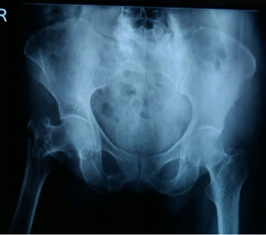
Radiograph of a right femoral neck fracture. Source: Wikimedia Commons — Drvaram; CC BY-SA 4.0. Image binary is embedded locally in this package; original source and licence are retained.

Talk
- I can see an AP view of left hip showing intracapsular fracture neck of femur. I would like to know the history especially the age, MOI and time since injury.
- In hight energy injury, I would approach using ATLS principles. I will first rule out any life threatening injury before looking for any limb threatening injury. I would also ensure that the injury is isolated closed and the limb is NV intact.
- I would like to see the lateral view and full length X-ray of femur and knee ap, lateral and skyline view. I will give adequate analgesia and a AK skin traction.
- I will arrange for an emergency table next day trauma list, as morning first case.
- After obtaining proper consent, marking and positioning the limb on a fracture table under adequate anaesthesia, WHO time out will be carried out. I will brief the team that first a closed reduction will be attempted by leadbetter techniques, and if the reduction seems inadequate, an open reduction using SP approach will be undertaken and fracture fixed with 3 6.5mm cannulated cancellous screws.
Key points
Reduction adequacy
- Gardens alignment index
- Lowells alignment theory
- Shentons line
Open reduction
- SP approach
- Plane: Femoral and SGN (Sart and TFL)
- Direct head of RF is released from AIIS
- Reflected head is released from capsule
Bhandari et al
- No significant difference in result
- Less reoperation with DHS
- 92% surgeons preferred Cannutated screw
| Column 1 | Column 2 | Column 3 | Column 4 |
|---|---|---|---|
| DHS | Cannulated screw | ||
| Invasive | More | Less | |
| Bone preserving | Less | More | |
| Rotational stability | No | Yes | |
| Mechanical strength | Better | Less |
Post op
- Toe touch for 6 weeks
- Radiological followup for 2 years
Complications
- Loss of reduction
- AVN
- Non union (powels valgus IT osteotomy with BG)