Talus
Clinical image
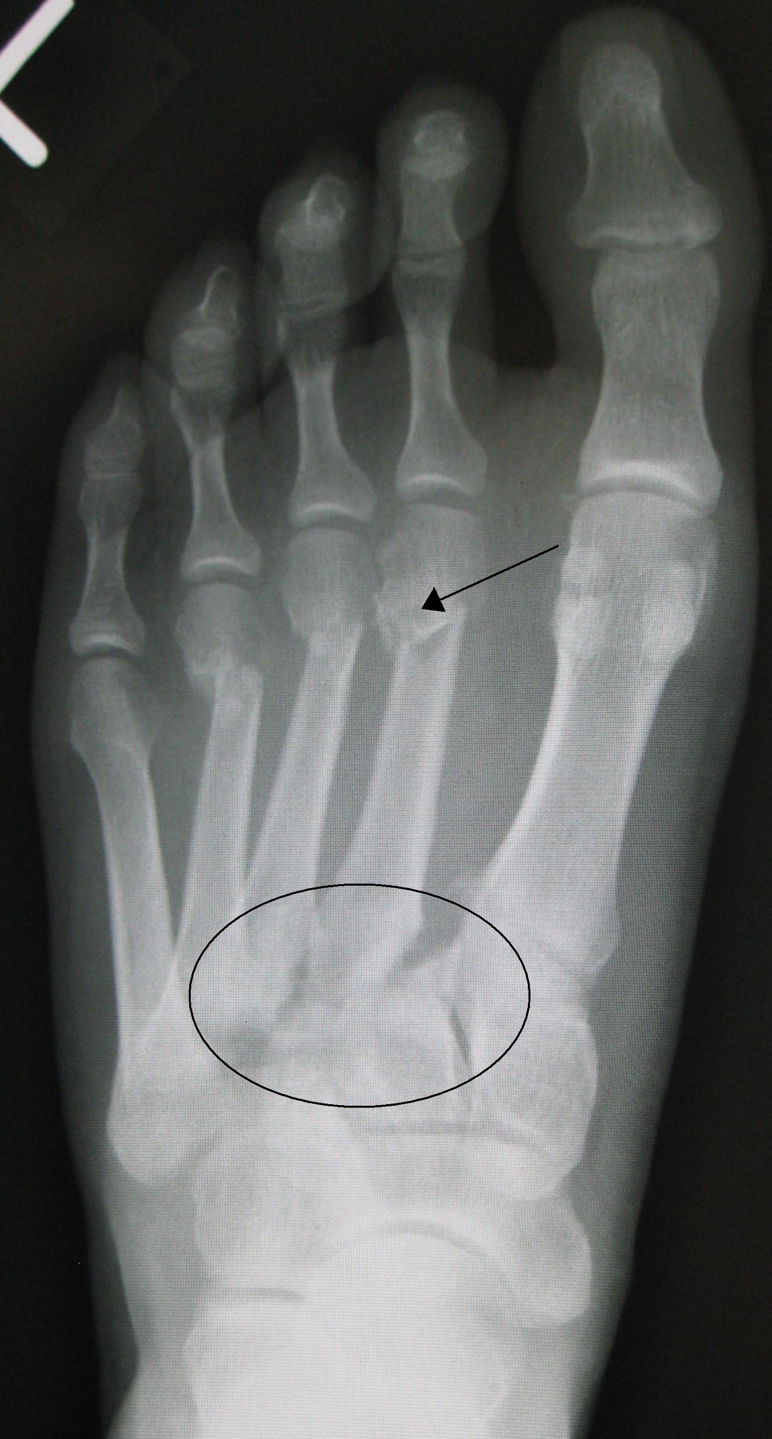
Radiograph of a Lisfranc fracture-dislocation. Source: Wikimedia Commons — James Heilman, MD; CC BY 3.0. Image binary is embedded locally in this package; original source and licence are retained.

Calcanium
- I see the lateral view of left ankle showing intra articular fracture of the calcaneum. I would like to know the MOI and time since injury
- I will approach patient using ATLS principles and ensure that the injury is isolated, closed, neurovascularly intact and there is no compartment syndrome, and assess the state of soft tissue. I will actively look for spine fracture and long bone fractures.
- I would like to see the axial view and bordens view.
- My management include
- Initial
- Planning
- Definitive
- Initially, I will give adequate analgesia and immobile in a back slab. Key is proper soft tissue care. That include cryotherapy, foot pump, anti-inflammatory medicines and limb elevation.
Talk
At this time I will order for a CT scan to know the pattern of injury to bone.
Comminution , widening of heel and lateral wall blow out all suggest operative fixation but my decision to operate will be based on
- Patient factors
- Soft tissue status
- Fracture pattern
Final decision will be taken in consultation with the patient
I will assess the patient for poor prognostic signs
- DM
- Smoking
- Male
- Manual labour
- Workers compensation
- Steroids
Patients who may benefit from surgical treatment include
- Gross deformity
- Open fracture
- Fracture causing soft tissue compromise
- Griffine et al BMJ 2014, RCT, no difference in outcome by 2 years
Principle
- Reduce CC and ST joints by reduction of Posterior facet and correction of widening of calcaneum
Compli
- Malunion (I: lateral exostosis, II: +ST arthritis,III: +varus)
- Impingement
- Compartment
- Wound healing
- FHL problem
Lisfranc
- 3 cuniforms and first 2 metatarsals
- Lisfranc ligament: runs from plantar second MT to plantar medial cuniform
- Fleck sign
X Ray
- AP: Medial border of 2nd MT in line with medial border of 2nd cuniform
- Oblique: Medial border of 4th and medial border of cuboid
- Lateral view : no step
Hardcastle
Treatment
- Screw or plate fixation of medial column and k wire fixation lateral column
- Suture anchor
- Arthrodesis
- Missed or failed ORIF
- Ly et al
- Crushed mid foot
- comminution
- multidirectional instability
- Mulier et al: Partial fusion