Clinical image
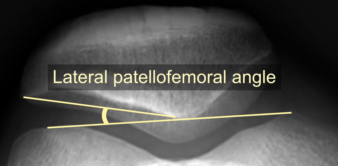
Axial patellofemoral radiograph demonstrating the lateral patellofemoral angle. Source: Wikimedia Commons — Mikael Häggström, M.D.; CC0 1.0. Image binary is embedded locally in this package; original source and licence are retained.

Knee Examination
PF Joint
- Stand: look for alignment, wasting, LLD
- Walk: Varus/ valgus trust, foot progression
- Sit: J Sign, Patellar height, Cripitus
- Lie down: Feel for tenderness and move
- Glide, Apprehension (In slight flexion), Clarke test
- Q angle
- NV status, Beighten score
Source of pain
- Black box of Orthopaedics
- Synovium, Subchondral bone or nerve
Causes
Local
Distant
Muscle/ soft tissue imbalance
- Tight Q,H,ITB,Lateral retinaculum
- Lax: BHES
Articular cartilage damage
- Trauma
- degeneration
- Inflammation
Bone stress
- OS disease, S-L J syndrome
Synovium: Plica
Fat: Fat pad syndrome
Causes Distant
- High q angle causes
- Femoral antiversion
- Valgum
- External tibial torsion
- Pronated foot
- Referred pain
Q angle
- Important in undestanding patellar malalignment
- Increased Q angle lead to patellar instability
J sign and Glide
- Distal realignment is beneficial
- Glide test positive: Lateral release
Patella Instability
Risk
- Static factors: Bony
- Hypoplastic femoral condyle
- Trochlea dysplasia
- Patella shape
- Patella alta
- Dynamic : soft tissue
- MPFL rupture
- Malalignment (> Q angle/ >TT-TG ratio)
- Abnormal gait
- Valgus trust
MPFL
- Primary soft tissue restraint
- Origin: between medial epicondyle and add tubercle (Schoulte point)
- Insertion: Prox 2/3 patella
- 5.5 cm long
- Torn in 90% acute dislocations
Treatment
- First line is always non surgical :
- Immobilise 4 weeks, rehabilitation
TT-TG distance
Surgical treatment
- MPFL reconstruction
- No other structural problem
- Proximal realignment
- Lateral release: rarely done in isolation
- Medial imbrication
- Quadriceps plasty
- Distal realignment (TT-TG >20mm)
- Medial transfer of TT in malalignment only (Elmslie Trillat)
- AM transfer if PF chondrosis also (Fulkerson)
- Distalisation if Insall ratio >1.3
PF arthritis
- Causes are same as that of Instability
- Radiology poorly correlates
- Management
- Non operative including I/A steroid
- Operative
- Arthroscopic debridement /microfracture
- AM transfer of TT
- PFA
- TKA
PFA
- Avon implant
- 10-14% revision rate in 8 years
- C/I
- Maltracking
- Instability
- Progressive disease pattern
- TF OA
- Chondrocalcinosis
Investigations
- Xray: Lateral and Merchant view
- CT
- MRI