Clinical image
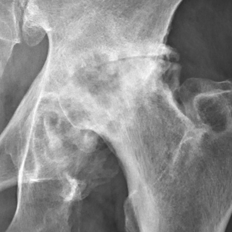
Severe hip osteoarthritis (Tönnis grade 3). Source: Wikimedia Commons — Mikael Häggström, M.D.; CC0 1.0. Image binary is embedded locally in this package; original source and licence are retained.

Key points
- 10% primary
- 90% secondary
- AVN
- DDH
- Perthes
- SUFE
- Protrusio
- Trauma
Management
- History: pain and functional limits
- Cl exams
- Investigate
- Assuming all conservative measures are tried and have been unsuccessful, I would offer her cemented THA
Which THA
- I will use a force closed taper slip cemented femoral stem of ODEP 10A rating
- Should have straight forward instrumentation
- Introducer should allow even pressurisation
- I will use a direct lateral approach
Hardinge
- Assuming that patient is adequately consented, relevant notes and X-rays are obtained, leg marked and prepared and WHO time out done
- Lateral position
- Landmark: ASIS, GT, Line of femur
- Incision: Longitudinal mid lateral 5 cm proximal and 10 cm distal to tip of GT
- IN plane: none
- 3cm above GT is Superior gluteal nerve
- Structures at risk: SGN, FNV, transverse br of circumflex femoral a
Posterior approach
- Double risk of sciatic nerve injury
- Similar time
- Less visible acetabulum
- Increased risk of cup anti version
- More dislocations
- Less Trendelenberg gait and better haris hip score
Posterior
- Assuming that patient is adequately consented, X-rays and notes are obtained, leg marked and prepped and WHO timeout done,
- my position of patient is: Lateral
- Landmark: GT
- Incision: with hip flexed 90 degree, a straight incision hand breadth below iliac crest on to the GT down to SOF.
- IN plane: none
- Superficial: split G Max
- Deep: Divide SER close to femur, except QF (Br of LSFA)
- Sciatic n: deep to piriformis and superficial to SER
- Risk: Sciatic n, Br of Inf Gluteal vessels
OA
- OA is a complex degenerative disorder of the entire synovial joint characterized by
- progressive loss of articular cartilage and
- subchondral bone remodelling.
Pathology
- Initial change is the damage to Tangential zone
- Disorganisation of collagen network
- Loss of proteoglycan
- Swelling
- Hypertrophic repair process starts
- Repair fails: leads to fibrillations parallel to surface
- Balance between anabolic and catabolic process is shifted in favour of catabolism
Key points
Pathology-Cartilage Catabolism
- Cartilage break down products are released in to synovial fluid causing inflammation and Macrophage activation
- Breakdown products
- Chondroitin so4
- keratan so4
- PG fragments
Macrophage
- PMN activation: Synovitis
- Release
- Cytokines
- Proteinases
- Oxygen free radicals
Cartilage changes
- increased
- water content
- chondrocyte activity and proliferation
- IL1
- MMP
- Cathepsin B and D
- Decreased
- Collagen content
- PG quality and size
- Keratan SO4
- Youngs modulus
MMP
- Molecule responsible for cartilage degradation
- Secreted by synoviocytes and chondrocytes
- Activated by IL1
- Include
- Stromelysins
- gelatinise
- collagenase
Histology
- Loss of chondrocytes
- Replication and Breakdown of tidemark
- Eburnation (Ivory like appearance) of subchondral bone