Clinical image
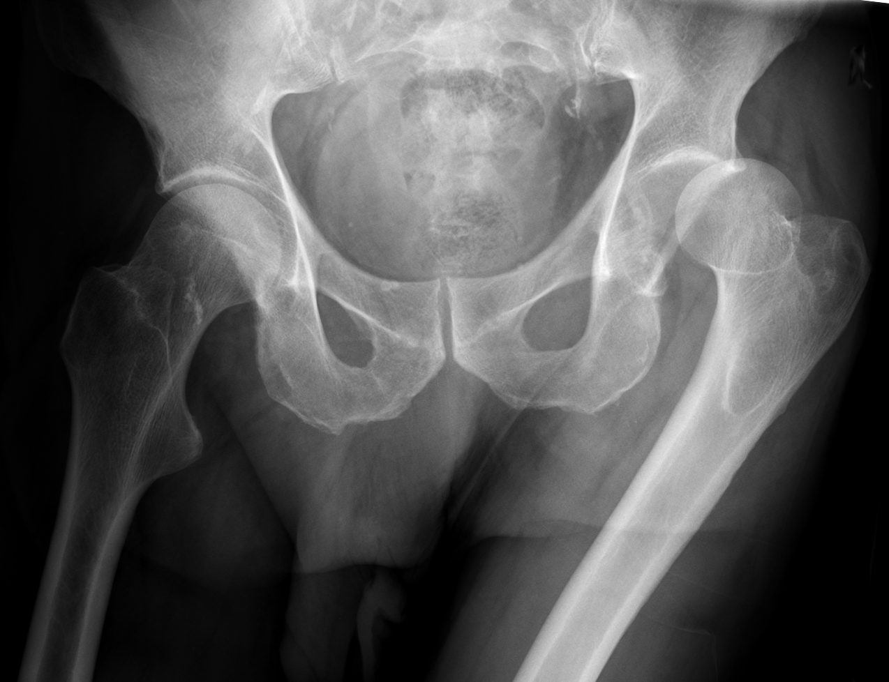
Radiograph of a left hip dislocation. Source: Wikimedia Commons — Hellerhoff; CC BY-SA 3.0. Image binary is embedded locally in this package; original source and licence are retained.

Key points
Talk
- I see AP view of pelvis with both hips with a fracture dislocation of the right hip. I will approach the patient using ATLS principles. After ruling out life threatening injuries, I will assess for the limb for neurovascular integrity, and exclude any open injury.
- I would like to see the lateral view and the entire length of femur, knee ap lateral and skyline view.
- This is a TE type v injury. I will give adequate analgesia and skin traction and arrange for emergency closed/open reduction of the hip. If CT can be taken without delay, that will help me to estimate the complexity of injury. Post reduction I will check for the stability and NV status
- I will also arrange for a post reduction CT