Clinical image
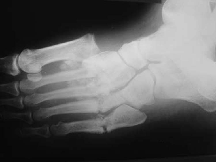
Charcot neuroarthropathy with Lisfranc disruption in a patient with diabetes. Source: Wikimedia Commons — J. Terrence Jose Jerome; CC BY 3.0. Image binary is embedded locally in this package; original source and licence are retained.

Foot ulcers
- precedes 80% amputations
- 1st amputation makes further amputation risk 23 times
- 70% death in 5 years after amputation
- 50% death after ulceration in 5 years
10% all hospital admission of DM
- 1/3 neuropathic
- 1/3 vascular ischemic
- 1/3 mixed
- MDT approach
Neuropathy
- Sensory:
- stocking destribution of sensory loss
- 10g Sammes-weinstein monofilament test: protective sensation
- Loss of vibration with 128hz early sign of neuropathy
- Motor
- Claw due to intrinsic weakness
- Equinous: weak Tib Ant
- Autonomic
- Dry skin
- altered nail growth
- reduced vascular response to injury
Assessment
- Diabetic control
- NV status
- Ulcers
- Deformity
Sammes Weinstein Monofilament
ABPI
Transcut oxygen pressure and angiography
NV status
Ulcers
- Neuropathic
- Painless under MT head
- Punched out with surrounding hyperkeratosis
- Good granulation
- Loss of protective sensation
- Normal pulses
- Ischemic
- Anywhere
- Painful
Deformity
- Claw toes
- Equinous
- Plantar keratosis
- Charcot asso deformities
- Cavus
- rocker bottom
Management
- Diabetes: control
- Ulcer
- Neuropathic
- accommodative foot wear
- Total contact cast
- Vascular
- MDT approach (angioplasty/bypass)
- Infected ulcer
- Step ladder approach from Ab to Amputation
- Neuropathic
- Deformity management
Deformity management Charcot
Goal of management
- Reduce deformity
- Provide stability
- Prevent ulceration
Charcot Deformity treatment principles
- DM control
- Accomodative foot wear
- TCC
- Avoid operations in destruction phase unless a/c deformity
- Arthrodesis preferred over ORIF
- Treat Equinous
- Long term non wt bearing (2-4months) followed by protective wt bearing for 1 year
- a/c deformity: surgery
- c/c deformity: non surgical
Operative indications
- a/c deformity
- Medial dislocation of navicular and cuniforms with foot in abduction, causing medial ulcerations: Osteotomy
- Dorsal dislocation of N and Cu causing lateral rocker bottom : Arthrodesis
- c/c progressive deformity
Emergencies
- a/c infection that does not require drainage
- a/c infection requiring drainage
- with out circulation compromise: drain by local facility
- with circulation compromise: Drain by vascular team
- Ischemic foot
- MDFS with in 24 hours
- Vascular input with in 48 hours
- revascularisation with in 8 days
Key points
- Summary of indications for conservative surgical approach or primary amputation
| Debridement/minor amputation | Primary amputation |
|---|---|
| Good blood supply to foot but infected | wet gangrene (infection + ischaemia) |
| Small vessel disease and gangrenous toes | life-threatening sepsis |
| Successful surgical bypass | extensive muscle necrosis |
| Neuropathic foot with little arterial disease | revascularisation technically impossible, bed-ridden patients/functionally useless limb |
| Osteomyelitis with little arterial disease |