Clinical image
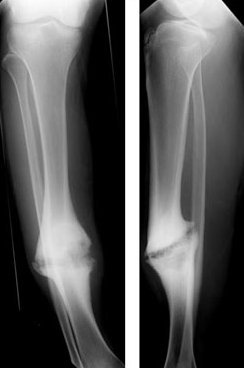
Hypertrophic tibial nonunion. Source: Wikimedia Commons — Lindsaydavidson; CC BY 3.0. Image binary is embedded locally in this package; original source and licence are retained.

- this X-ray?
Non union
Describe what you see
- I see the lateral view of a right femur with an ante-grade IM nail used to fix a diaphysial fracture. What concerns me is the atrophic non union.
- Ask for other views
Definition
- Biological process of fracture healing has come to a stand still which is evident clinically and radiologically.
- Causes of non union are multiple and my management will be based on the causes which I divide in to
- Infection
- Local cause
- Systemic cause
- Mechanical
Local causes-infection
- Any non union is infected unless proved otherwise
- I would assume that there are no sinuses, wounds healed by secondary intention, history or chronology of events suggestive of infection. How ever I would exclude infection by blood markers and culture of tissue samples next to bone.Mri, leukocyte scan are useful adjutants .
- mri is most sensitive, but less specific. leukocyte scan is good for acute infection, but in c/c rumbling infection they are less useful.
- Cierny and mader classification
Key points
Local factors
- Index injury
- Fracture personality gustilo etal
- open fracture
- compartment syndrome blair
- soft tissue injury Tscherne
- Surgical technique
- Tissue handling
- tourniquet
- wound coverage
- approach
- distraction
- reamers
- appropriate application of fixator/cast
Systemic
- Patient co morbidities
- DM
- COPD
- PVD
- Steroids
- Endocrinopathies
- VD and V K
- Patient social
- Alcohol
- smoking
- non compliance
Mechanics
- Principle applied
- was it appropriate? achieved?
- Stability
Management
- This is a difficult problem
- Assess the causes and address the causes
- MDT approach
- Confer with colleagues with more experience
- How ever my goals are
- To achieve union
- eradicate any infection
- address shortening/malalignment
- Achieve good function
Management Planning
- Host type
- soft tissue status and coordination with plastic surgeon
- Bone factors-shortening/alignment/bone loss/joint stiffness
Septic non union
- 3 stage procedure
- First stage- remove implant, resect devitalised bone and soft tissue, local/systemic antibiotics, temporising ex fix. Serial blood markers, blood culture, repeated debridement if needed, VAC
- Second stage- Soft tissue coverage by plastic
- Third stage- Definitive procedure, nail/plate/circular frame
Hypertrophic no previous surgery
- If in good alignment and no shortening/gap
- Functional cast/ LIPUS (Low intensity pulsed Usg)
- Nail/plate fixation
Atropic no previous Sx
- Associated gap, shortening and malalignment
- Circular frame
Aseptic with previous Sx
- Single stage revision fixation with graft/ BMP after actively ruling out infection