Anatomy
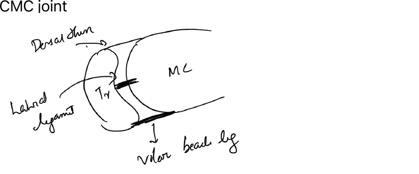
The first carpometacarpal joint is a saddle joint between the base of the first metacarpal and the trapezium. Its mobility permits thumb opposition but also makes it susceptible to instability and osteoarthritis.
Important stabilising structures include the dorsoradial ligament and volar/anterior oblique ligament complex.
Basal thumb arthritis
Degeneration of the trapeziometacarpal joint causes:
- Pain at the base of the thumb
- Loss of pinch strength
- Crepitus
- Progressive subluxation and adduction deformity
Examination
- Tenderness over the CMC joint
- Painful grind test
- Pain with key pinch
- Assess MCP hyperextension
- Assess first-web-space contracture
Imaging
Standard hand or dedicated thumb radiographs may show:
- Joint-space narrowing
- Osteophytes
- Subchondral sclerosis
- Subluxation
- Scaphotrapeziotrapezoid involvement in advanced disease
Non-operative treatment
- Activity modification
- Thumb-spica splint
- Analgesia
- Hand therapy
- Intra-articular corticosteroid injection in selected patients
Operative treatment
Options depend on age, demand and disease stage:
- Trapeziectomy, with or without ligament reconstruction / tendon interposition
- Suspension procedures
- Arthrodesis in selected younger high-demand patients
- Implant arthroplasty in selected cases
MCP hyperextension may need simultaneous correction when substantial.
References
- Orthobullets. Basilar Thumb Arthritis.