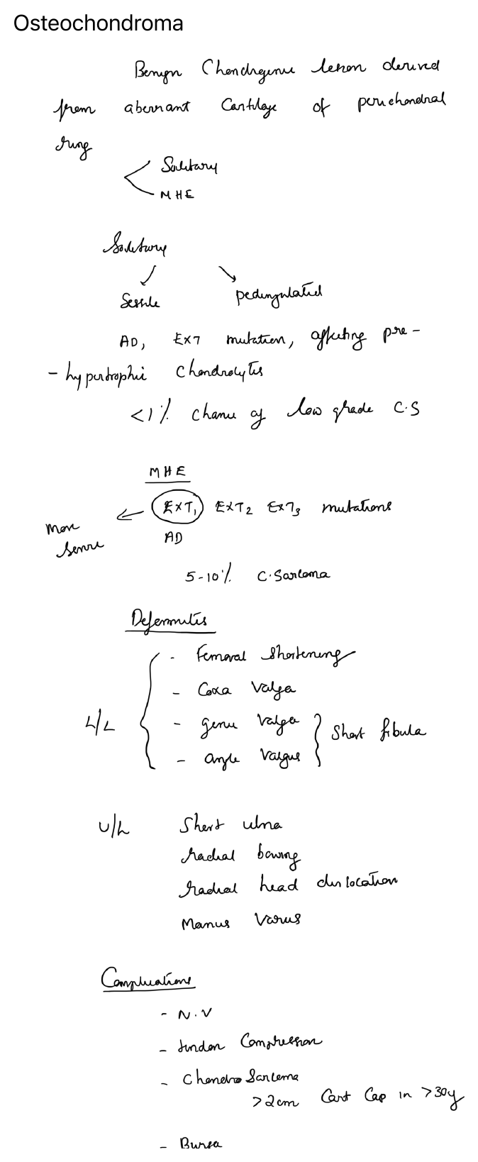
Definition
Osteochondroma is a cartilage-capped bony exostosis arising on the external surface of bone and continuous with the cortex and medullary canal of the parent bone. It is the commonest benign bone tumour.
Types
- Solitary osteochondroma
- Multiple hereditary exostoses (MHE)
Usual sites
Typically occurs in the metaphysis of long bones, especially around the knee:
- distal femur
- proximal tibia
- proximal humerus
Clinical features
- Painless lump
- Mechanical symptoms from tendon or muscle irritation
- Cosmetic concern
- Symptoms due to pressure on adjacent structures
Imaging
Plain radiographs
- Sessile or pedunculated bony outgrowth
- Cortex and medullary canal are continuous with those of the host bone
MRI
Useful when malignant change is suspected or when the relation to neurovascular structures is important.
Complications
- Bursitis over the lesion
- Mechanical irritation
- Fracture of a pedunculated stalk
- Neurovascular compression
- Deformity in multiple hereditary exostoses
- Rare malignant transformation to secondary chondrosarcoma
Suspicion of malignant transformation
Consider if there is:
- new pain
- increase in size after skeletal maturity
- cartilage cap thickening
- irregular or destructive imaging features
Treatment
- Observation for asymptomatic lesions
- Excision for pain, mechanical symptoms, deformity, neurovascular compromise or suspicion of malignant change
Diagram