Extracapsular
Clinical image
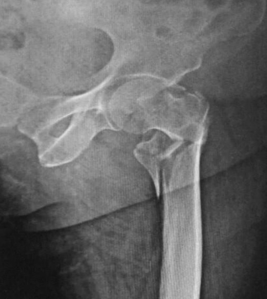
Radiograph of a comminuted proximal femoral fracture. Source: Wikimedia Commons — Rohan R. Memon, Drashtant Patel, Nishant Juva; CC BY 4.0. Image binary is embedded locally in this package; original source and licence are retained.

Key points
Unstable fracture patterns include:
- • Reverse oblique
- • Transtrochanteric
- • With subtrochanteric extension
- • With ‘large’ posteromedial fragment – although the “large” is not well defined
Transtochantric
Reverse oblique
Posteromedial comminution
Key points
| Column 1 | Column 2 | Column 3 | Column 4 |
|---|---|---|---|
| DHS | Nail | Trochanteric stabilising plate | |
| Unstable # | No | Yes | |
| Lateral wall comminution | No | Yes | Yes |
Predictors of failure
- Boumgartner TAD >25mm
- Huang Lateral wall <20.5