Clinical image
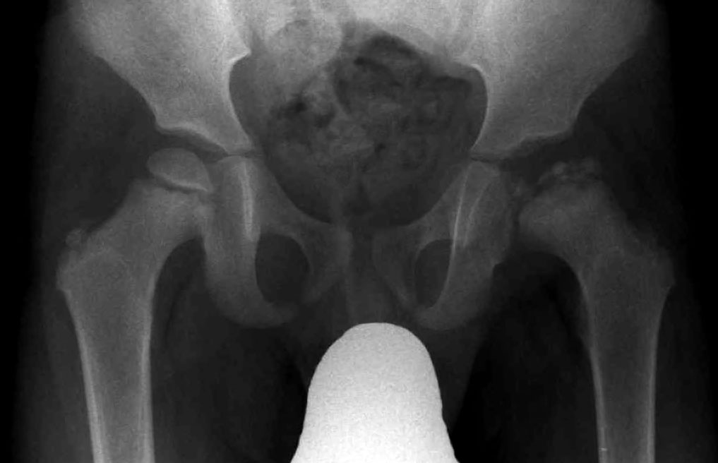
Radiograph showing Legg–Calvé–Perthes disease. Source: Wikimedia Commons — J. Lengerke; Public domain. Image binary is embedded locally in this package; original source and licence are retained.

Key points
AP X-ray of pelvis shows flattening and sclerosis of right femoral head. Acetabulum appears normal. I would want to exclude infection, but my primary diagnosis is Perthes disease
Talking the talk
- It is a self limiting disease of children characterised by reversible interruption of the blood supply to CFE resulting in necrosis of epiphysis.
Pathophysiology
- Bone necrosis triggers healing response
- This triggers synovitis,cartilage and Lig teres hypertrophy
- Soft tissue changes and muscle spasm causes femoral head extrusion
- Extrusion is the primary cause of deformation
Elizabeth Town
- 1: Avascular necrosis: sclerosis
- 2: Fragmentation
- 3: Reossification
- 4: Healed
- a and b based on early or late
- Any treatment done after 2b is only remedial or salvage
Management of Perthes D is controversial. I will discuss with Paed Orthopaedic surgeon.
Principle of treatment is
A rationale approach of treatment to prevent/minimise femoral head deformity in the early course of disease
A rationale approach of treatment for late presentation
Containment
- Done in early in the course of disease based on age
- It is placing the A-L part of femoral head well in to the acetabulum
- Methods:
- Keep hip abducted and IR/Abd and flexed by
- Cast
- Brace
- Surgery on femur
- Acetabulum reoriented to cover A-L femoral head
- Salter
- shelf
- Keep hip abducted and IR/Abd and flexed by
Age, stage, stiffness and extrusion
- Odds of avoiding deformation 16.58 if contained before 2b stage
- ROM should be regained before containment by traction
- If ROM not improving, give broomstick cast for 6 weeks
| Age | Stage | Extrusion | Treatment |
|---|---|---|---|
| >8 years | <2b | NA | Contain |
| <8years | <2b | Yes | Contain |
| <8 years | <2b | no | Followup 3 mo for extrusion |
VDO
- Kim 10-15 degree
- Benjamin Joseph 20 degree
Two factors widely used for treatment planning, the status of the lateral pillar (Herring's grading) and the extent of epiphyseal involvement (Catterall's grouping) have not been included in the decision-making scheme.
This is because treatment needs to be initiated well before the stage of fragmentation when these two variables can be identified with some degree of certainty
Late in the course =>2b
- Remedial surgery: head has already deformed permanently
- Hinged abduction: Valgus Ext Osteotomy
- Extrusion: shelf
- Promote healing: arthrodiastasis and epiphyseal drilling
Treatment of sequelae (salvage)
- Joint preserving surgery if no OA
- If OA: THA (has reduced Euroquol)
Etiology
- Vascular
- Thrombophilic theory
- Trauma
- Passive smoking
- Genetic
- Susceptibility
- LBW
- Low socioeconomic
- delayed bone maturation
- Boys> girls
C/E
- FADER
- Trendelenberg positive
- LLD
- Height and exclude dysplasia
b/l dd
- MED
- Hypothy
- SED
- Meyers dysplasia
- MPS
- Sickle
Xray
- Widened joint space
- Small CFE
- Cresent sign
- Flat epiphysis
Head at risk signs
- Clinical
- Obesity
- Decreased ROM
- Adduction contracture
- Radiological
- Calcification lateral to physis
- Metaphysial cyst
- Increased medial space
- Horizontal physis
- Gage sign (v shaped lucency lateral to physis)
Lateral Piller
- Group A: No lateral filler involvement
- Group B: > 50% height involved
- Group C: < 50% height maintained
- Predicts likelihood of containment
Salter Thompson
- Group A: <50% head involved
- Group B: >50%
Catteral
- Group 1: Anterior ep involvement
- Group 2:Central collapse
- Group 3: Head with in head
- Group 4: Whole head involved
Stulberg
- Spherical congruent
- Aspherical congruent
- Aspherical non congruent