Clinical image
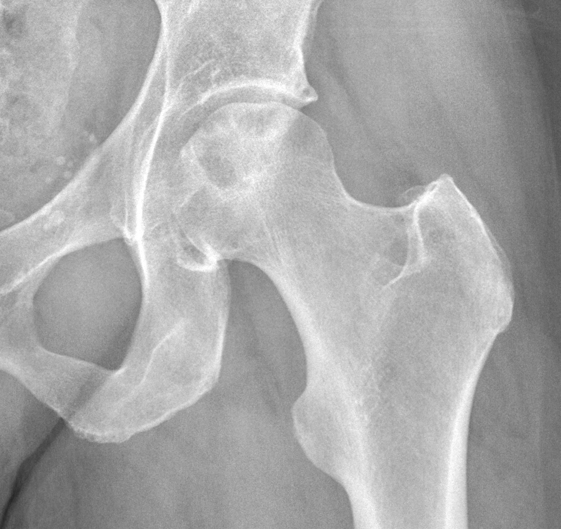
AP radiograph of avascular necrosis of the femoral head. Source: Wikimedia Commons — Mikael Häggström, M.D.; CC0 1.0. Image binary is embedded locally in this package; original source and licence are retained.

Key points
- AP view of pelvis with both hips showing diffuse sclerosis with increased density in the superolateral aspect of both hips suggestive of AVN. I would like to see frog leg lateral view
- To see crescent sign suggestive of subchondral fracture
AVN
- Osteonecrosis of the femoral head due to interruption of blood supply leading to ischaema and cell death
- Etiology
- Steroid
- Smoking
- Alcohol
- Idiopathic
AS IT GRIPS 3 C
- Alcohol
- Steroid
- Idiopathic
- Trauma
- Gaucher
- Rheumatoid
- Infection
- Pregnancy
- Smoking
- CRS/Chemo/Caisson
Pathology
- Related to underlying ethiology that alters the blood flow
- Five vascular areas that can be involved are
- Extra osseous arterial most important
- Trauma/vasculitis/spasm
- Intra osseous Arterial due to circulating micro emboli
- Fat embolism/Caesson/SCD
- Extra osseous extravascular
- Capsular tamponade
- Intra osseous extravascular
- Fat cell hypertrophy (steroid)
- Abnormal cells (Goucher)
- Intera osseous venous
- stasis in SCD/Fat cells
- Extra osseous arterial most important
- Clotting abnormalities
Steroid AVN
- Fat cell hypertrophy
- Increased fat cell content
- Fat embolism
- Endothelial and smooth muscle cell damage
- Primary osteocyte death
- 10-30% of AVN, m/c atraumatic cause
2g prednisone > 2 months
Classification
- Ficat and Arlet with hungerfold modification
- 0: preclinical: Suspected disease in C/L hip
- 1: Pre radiographic: MRI shows double line sign/bone scan
- 2: Precollapse : Osteopenia, Sclerosis, cyst and cresent sign
- 3: Collapse: Flat head
- 4: OA with deformed head
Steinberg
7 stages
0: Histology
1:abnormal bone scan/MRI
2:Sclerosis, cyst
3:Cresent sign with out flattening
4: Flattening
5: OA
6:Advance OA
<15%
15-30%
30%
Others
- ARCO: Association research Circulation Osseous)
- University of pennsylvania
- Mitchell MRI classification
Kerboull Necrotic angle
- Necrotic angle is measured in AP and Frog lateral
200 degree: poor result with bone preservation
Management
- Goal is to
- relieve pain
- prevent deformity
- preserve function
- Based on
- Precollapse or not
- Necrotic segment
- Femoral head depression
- Acetabular involvement
Non surgical
- Collapse occurs in 80% patients with pain with in 4 years
- Pharmacological
- Alendronate: Agarvala et al , JBJS 2009, 2% failure in 1, 8% in 2 and 33% in stage 3
- Iloprost: vasodilator
- Statins: pro osteoblastic and anti adipogenic
- Electrical stimulation: experimental
- Hyperbaric O2: reversal of cellular ischemia
Stage 1 and 2
- Core decompression: relieve pressure, increase vascularity and healing
- 2/3 good result (Mont et al)
- Tantalum rod insertion: no donor site morb, structural supp, osteointegration
- 15 % failure rate
- VFG/non VFG
- Medical: Bisphosphonates, Anticoagulants, Vasodilators, Lipid lowering agents
- Stem cells: one stage or two stage
- Trapdoor
- Muscle pedicle BG
VFG
- McKee indications
- <45
- 2mm or less collapse
- etiology withdrawn
- no contracture
- joint supple
- Advantages: support subchondral bone and provide blood supply for revascularistion
- compli: Gaskill
- Donor site: Great toe contracture , weakness, pain sensory deficit
- Graft site: Pin migration, HO, Infection, Fracture, DVT
2 and 3
- Trapdoor: Mont et al 73% excellent result in 5 years
- Osteotomy
- Attempts to shift the involved part medially
- Angular: varus/valgus
- Rotational: Sugioka
- Indications
- Young <45
- Kerboull <200
- No joint narrowing
- Unilateral
- Traumatic/idiopathic
Arthroplasty
- Bipolar Hemi: elderly
- Hybrid THA
- Uncemented THA:
- Cheung et al in Hip Int, 2015: 117 AVN, age 51 mean, >95 % survival in 19 years
- SCD, Goucher, CKD, Trauma, NOF # higher revision rate
- Cemented
- Historically poor result
- Steinberg 96% survival with 2nd gen cementing
- Kim: No difference in result with hybrid and uncemented
Choice of THA
- < 55 years
- Uncemented THA CoC
- 6.35% revision in 10 year MoP
- 4.5% revision for CoP
- 3.5% revision for CoC
- Cemented THA CoP
- 7.5 % revision MoP
- 3.5% revision CoP
- Non controversial answer (Kim comparison, Steinberg)
- Cemented Forced closed Femoral stem with at least 2nd gen cementing
- Ceramic head
- Highly cross linked poly
- Uncemented THA CoC
CoC
- Hard, wear resistant and wettable
- Better lubrication
- Decreased wear
- Catastrophic Fracture, Squeaking, chipping during insertion
Asymtomatic ON
- Necrosis in MRI Kim et al
- Small (<30% head involved): 5% disease progression
- Medium (30-50%): 45%
- Large(>50%):85%