Clinical image
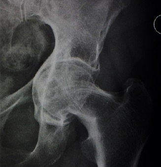
Radiographic appearance of protrusio acetabuli. Source: Wikimedia Commons — Fernando Ruiz Santiago et al.; CC BY 4.0. Image binary is embedded locally in this package; original source and licence are retained.

Key points
Hirst
5mm
10mm
15mm
- or CE angle >40 degree
Aethiology
- Decreased bone density
- OI
- Osteomalasia
- Osteoporosis
- Rheumatoid
- Marfan
- A Spondylitis
- Normal density
- Otto
- OA
- Increased density
- Paget
Management
- Exclude causes
- Xray
- CT for bone defect
- MR arthrogram if preservation planned
Preservation
- Valgus ITO and triradiate closure: young
- Periacetabular osteotomy
Principles of THA
- Assess bone defect
- Template
- Sciatic nerve is closer to hip
- Difficult dislocation
- Bone graft the floor and lateralise the cup, use uncemented THA
- Baghdadi et al, 90% 15 year survival