Clinical image
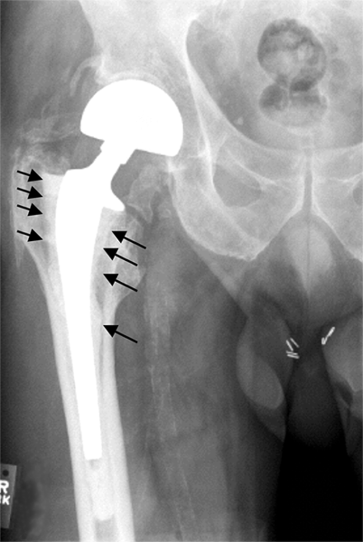
Peri-implant osteolysis in aseptic loosening before revision THA. Source: Wikimedia Commons — Zhenxin Shen, Tania N Crotti, Kevin P McHugh, Kenichiro Matsuzaki, Ellen M Gravallese, Benjamin E Bierbaum, Steven R Goldring; CC BY 2.0. Image binary is embedded locally in this package; original source and licence are retained.

Key points
Talking the talk
- AP radiograph shows osteolysis of the femoral component.. There are radiolucency lines at the bone cement interface located circumferential along all seven gruen zones.
- Femoral component is a Stanmore prosthesis and no cement plug is seen in X-ray, suggesting a first generation cementing technique.
Cementing techniques
Gruen zones
- Charnley and DeLee zones
Grading of cementing quality
Pain ful THA
- I will first exclude infection
- Causes can be extrinsic or intrinsic
- Exclude
- Referred pain
- Soft tissue causes: psoas/abductor tendinosis, impingement
- POVD
- Stress fracture
- Component malposition
- Femoral neck fracture
- Aseptic loosening
- Heterotropic ossification
What will you do?
- History
- Pain: intrinsic or extrinsic
- infection
- Examination
- Investigations
Offer revision
- Complications
- ETO, hence
- prolonged procedure
- more blood lose and
- osteotomy complications
- Assess bone loss: Paprosky
Planning the surgery
- Anaesthetic fitness assessment
- 4 units crossmatched blood
- Allograft: femoral head frozen
- Implant removal kit
- Templating
- Dall miles cables for ETO
- Flexible light source
- Posterior approach
Prefer uncemented implant
- Long stem femoral component
- Tantalum acetabular shell
- MoP
- at least 32 mm head
Cement removal
- Femoral
- Remove first from shoulder
- Flexible osteotome and burr
- ETO
- Acetabular
- First remove PE cup from cement
- Remove cement piecemeal