Clinical image
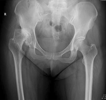
Adult developmental dysplasia of the hip. Source: Wikimedia Commons — Ping Zhen, Jun Liu, Hao Lu, Hui Chen, Xusheng Li, Shenghu Zho; CC BY 4.0. Image binary is embedded locally in this package; original source and licence are retained.

Key points
Talking the talk
- AP xray of the pelvis shows dysplasia of the left hip. The femoral head is subluxated but articulating with the true acetabulum, suggesting dysplasia. Head is deformed and there is secondary osteoarthritic changes. Proximal femoral canal appears narrow.
Dysplasia
- Lack of coverage of femoral head whether it is subluxated or dislocated
Classifications
Anatomical issues
- Soft tissue
- Capsule elongated
- Psoas hypertrophy
- Horizontal orientation of abductors
- Short femoral and sciatic nerve
- Femoral nerve lateral
- Acetabular
- Small shallow
- Femoral
- Trochanter small and posterior
- Narrow femoral canal
- Increased antiversion
CT
- Helps to know the bone stock, morphology, orientation and dimensions of both acetabulum and femur
Placement of acetabulum
- Anatomical
- Less loosening (Linde et al)
- LLD can be addressed
- Needs bone grafting/cotyloplasty
- Difficult
- May require femoral shortening
- High hip centre
- Does not require shortening of femur
- 3 times more loosening
- LLD cannot be addressed
- Bone stock is not preserved for revision
- High rate of instability
Femoral side
- Anteversion
- femoral fracture
- IR contracture of hip
- Increased instability
- modular implant may be needed
- Rotational osteotomy may be needed
- Narrow femoral canal: custom prosthesis
- Femoral shortening
- Sequential proximal resection
- Unsuitable for uncemented
- Subtr osteotomy
- Increased nonunion
- Sequential proximal resection