Clinical image
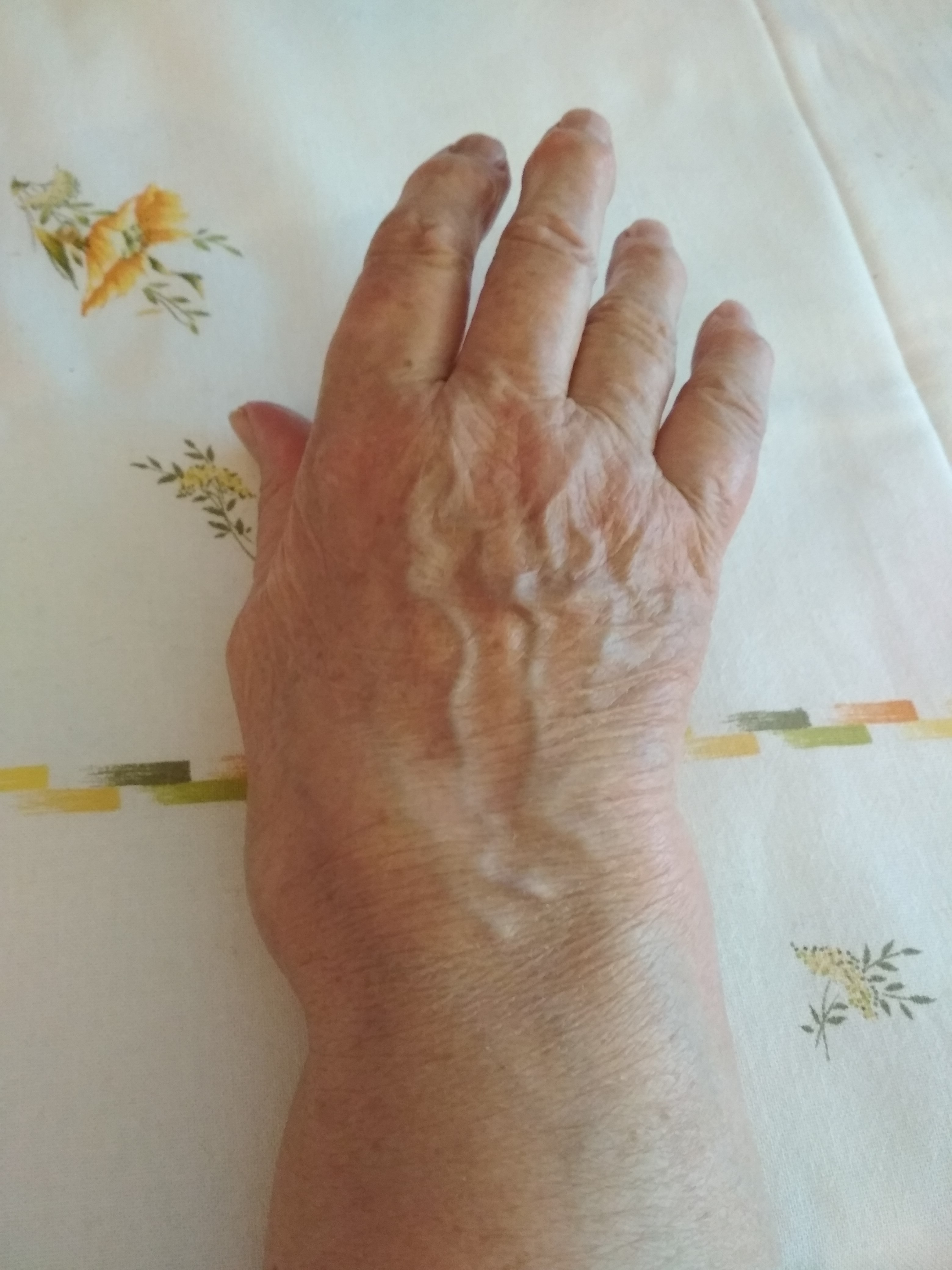
Clinical appearance of a hand affected by rheumatoid arthritis. Source: Wikimedia Commons — Amm0nium; CC BY-SA 4.0. Image binary is embedded locally in this package; original source and licence are retained.

Problems
- Pancarpal disease leading to fusion/palmar dislocation of RC articulation
- Periscaphoid: DISI
- DRUJ instability
- MCP joint disease
- Ext tenosynovitis
- Flexar tenosynovitis
- tendon rupture
- Trigger
- CTS
- Thumb
- Swan and bout
Wrist
- Volar subluxation of carpus
- Volar carpal lig laxity
- Caput ulna leading to vaughan jackson
- Volar subluxation of carpus
- ECU subluxation
- DRUJ disease
Metacarpals and MCPJ
- Radial deviation
- Volar ulnar subluxation of MCPJ
- Altered pull of EDC
- Capsular laxity
Intrinsic imbalance
- Boutonniere
- Swan neck
- Ulnar deviation of fingers
- Radial collateral lig weakness causing volar ext tendon subluxation
Dropped fingers
- PIN
- Tendon rupture
- Tendon subluxation
Inability to flex DIP
- FDP rupture
- FDP FDS adhesion
- Trigger
Assessment
- Look:
- Screen neck, shoulder and elbow
- Describe deformities
- Swellings
- Scars
- Muscle wasting
- Move active
- Feel: Swellings
- Functional assesment
Functional assessment
- Power grip
- Tripod: Pen
- Key
- Precision: Coin
Rheumatoid surgery
- Lower limb first
- Forefoot first if hind foot flexible
- Hind foot first if not flexible
- Upper limb proximal first then distal
- Urgent procedures
- Nerve compression
- incipient tendon rupture
Common procedures
- Wrist arthrodesis (arthroplasty)
- DRUJ fusion/ excision
- MCPJ arthroplasty
- Thumb MCPJ arthrodesis
Wrist arthroplasty
- Prerequisites
- Ext tendons intact
- Minimal deformity
- Adequate bone stock
- No infection
- Other side arthrodesis
Reconstruction tendon rupture
| Problem | Procedure |
|---|---|
| FDS | None |
| FDP wrist | Tenodesis |
| FDP finger | DIP fusion |
| FDS and FDP | FDS other finger |
| FPL | IP fusion |
Vaughen Jackson
- Darrach + synovectomy for pre rupture
- Little F: suture to RF
- LF and RF: suture to MF
- MF, RF and LF:
- EI to RF and LF
- MF to IF
- All
- FCR transfer
Mannerfelt
- FPL
- Tenosynovitis of Scaphotrapezial region
Rheumatoid thumb
Key points
| Column 1 | Column 2 | Column 3 | Column 4 | Column 5 |
|---|---|---|---|---|
| Nalebuff | Problem | Location | Treatment | |
| 1 | Boutonniere | MCPJ extensor hood attenuation | Flexed MCP, extended IP | MCP fusion |
| 2 | 1+ CMCJ dislocation | MCP fusion | ||
| 3 | Swan neck | CMCJ disease | Subluxated CMC; Adducted thumb MC; Extended MCP compensation; IPJ flexion | Trapezectomy and MCP fusion |
| 4 | Gamekeeper thumb | UCL rupture | Adduction contracture | MCP fusion |
| 5 | MCPJ hyperextension | Volar plate | MCP fusion |
Boutonniere finger
Pathology
- PIP joint extensor aspect
- Attenuation of central slip
- Disruption of triangular lig
- Volar subluxation of lateral band
Classification Nalebuff
- Mild: <15 degree ext lag correctable (Splint)
- Moderate : > 15 degree lag with some contracture (Central slip reconstruction)
- Severe: FFD (Arthrodesis)
Capner splint for boutonnière
Swan
Pathology
- Imbalance of forces at PIP joint or lax volar plate leading to loss of flexion at PIPJ , hyperextension at PIPJ and Flexion at DIPJ