Clinical image
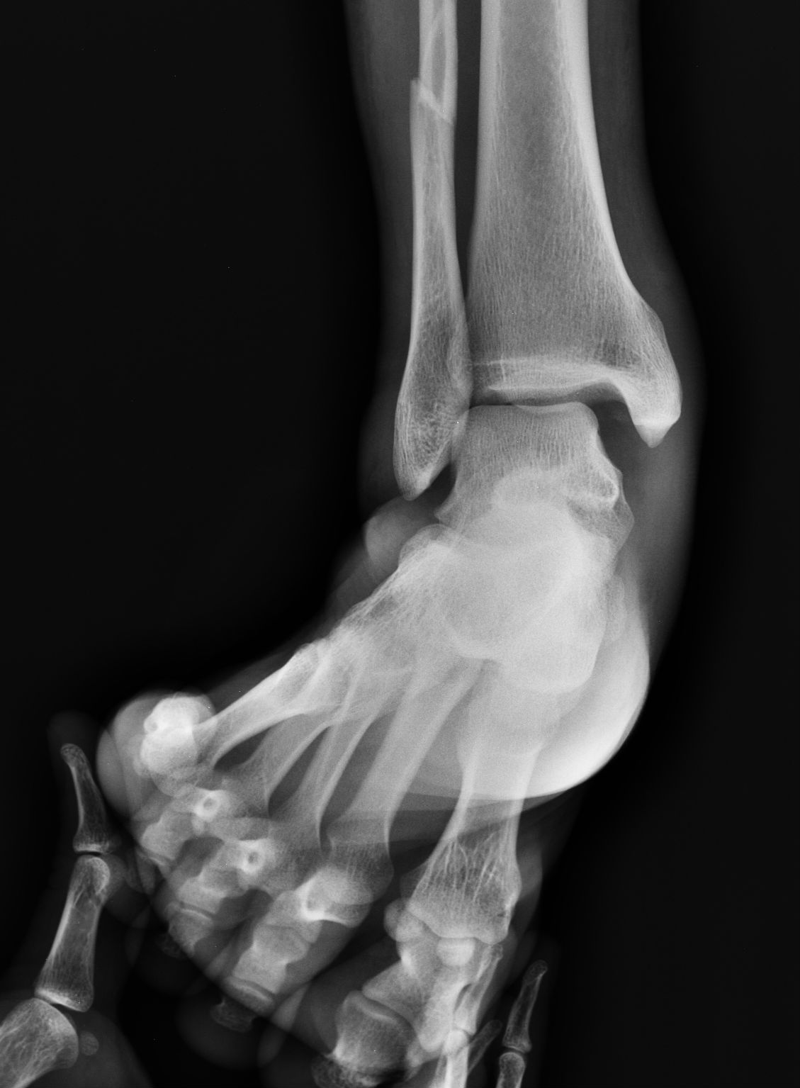
Ankle stress radiograph demonstrating instability. Source: Wikimedia Commons — Case courtesy of Dr Maulik S Patel; Commons upload Patellaton; CC BY-SA 4.0. Image binary is embedded locally in this package; original source and licence are retained.

- “Stress xray of ankle shows significant taller tilt, if associated with significant history and examination findings, this suggests ankle instability, probable chronic. This my be an indication for considering surgical management. That include anatomical and non anatomical repair. If there is significant risk factors such as high demand, hyper laxity and cavus, I will consider non anatomical repair and otherwise anatomical.
- A brace is given for 3 months and advice to continue to use a tape or brace indefinitely if the patient is involved in athletic activities ”
Ankle instability
Key points
Injury
- Stable
- Unstable
- Partial: Anterior drawer positive
- Complete: Anterior drawer positive and talus tilted
History
- A/C ligament injury 4-6 week conservative treatment
- Locking/Clicking: OC fragments, arthritis
- C/C Painless giving way: Ligament
- C/C Painful mechanical giving way: not ligament
- Impingement
- OC fracture
- Peroneal injury
- Arthritis
- Painful functional giving way:
- Internal impingement: EUA, Arthroscopy
So ask
- Giving way
- Duration
- Pain
- Mechanical/ functional
- Locking/clicking
- Functional demand
Examination
- Beighton score
- Hindfoot varus/ forefoot pronation and subtle cavus: m/c cause of recurrent instability
Management
- Xray :
- Talar tilt> 15 degree
- Anterior translation >3mm
- RICE and early functional rehabilitation
- 80% g3 injury can be treated non surgically with good result
- If not responding: MRI
Indications for surgery
- Failed conservative treatment
- c/c instability with anterior drawer and stress radiograph positive
Treatment
- Anatomical repair
- Non anatomical repair using tenodesis
- Anatomical reconstruction with tenodesis
Anatomical repair
- Restores normal kinematics
- Local tissue may be scared and suboptimal for the purpose
- No risk factors to be present
- Brostrom Gould
- Brostrom
- Direct repair ATFL/CFL
- Gould
- Augment with inferior retinaculum
Non anatomical repair
- Tenodesis is used to restrict motion
- Donot accurately restore kinematics
- Evans: peroneous brevis
- Chrisman snook: anterior half of PB
- Used if risk factors are present
- High demand
- Cavovarus
- Hyper laxity