Clinical image
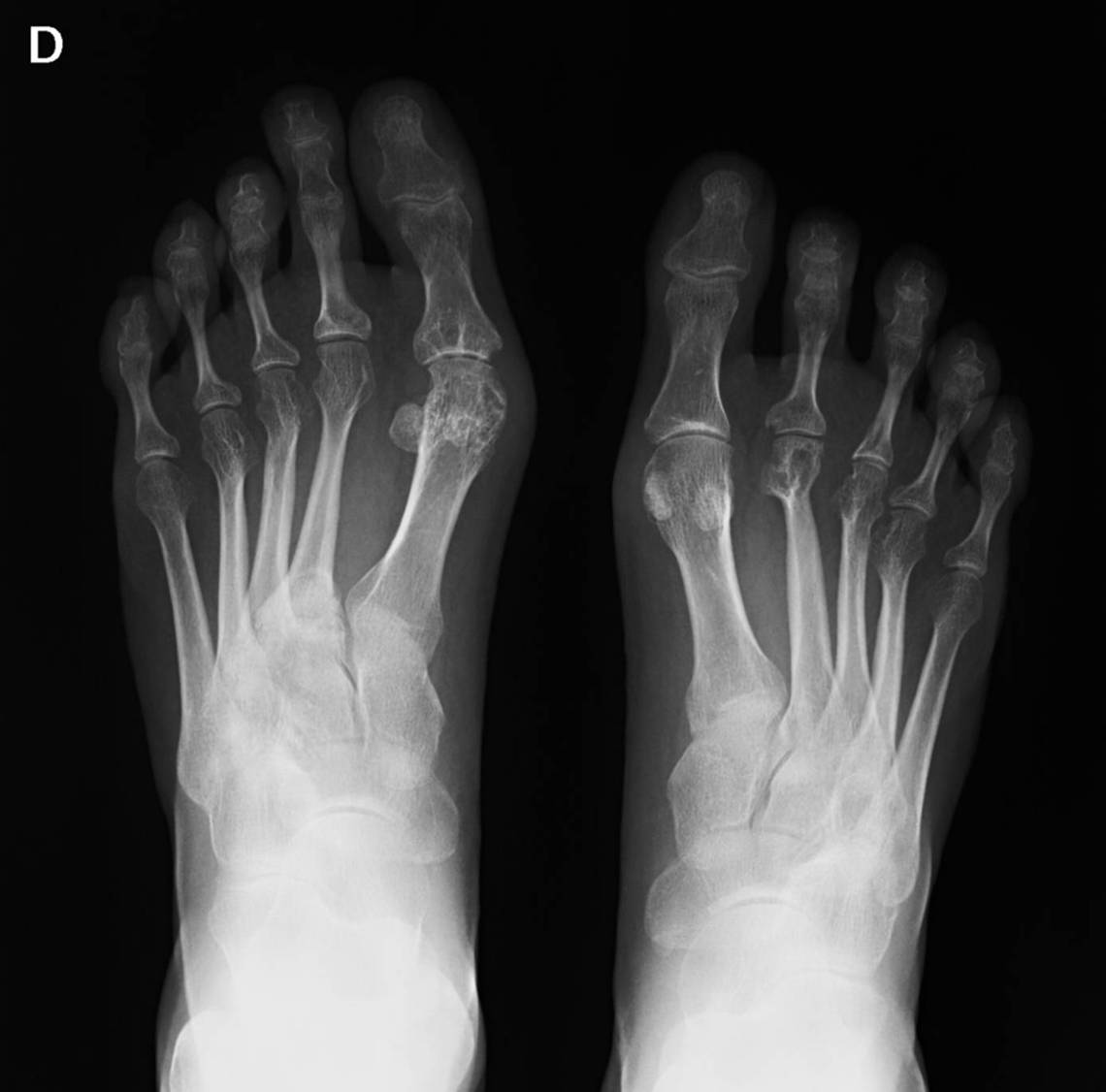
Radiograph of feet affected by rheumatoid arthritis. Source: Wikimedia Commons — Lariob; CC BY-SA 4.0. Image binary is embedded locally in this package; original source and licence are retained.

Key points
Deformities
- Forefoot
- Hind foot
- Soft tissue
Fore foot
- Hallus valgus
- 1st MTP joint erosive arthritis
- Dorsal sub/dis of MTP joint (Synovitis, plantar plate laxity)
- Prominent MT head in the sole (callosities)
- Hammer and claw toes (intrinsic contracture)
Hind foot
- Valgus ankle and instability
- Valgus hind foot due to subtalar arthritis
- Medial arch collapse and foot pronation (TN subluxation)
- Subfibular impingement
Soft tissue
- Tenosynovitis TP and Peroneals: ankle swelling
- Medial arch collapse: TP rupture and TC ligament
- Distal migration of fore foot fat pad
- TT syndrome
- Morton neuroma
- Retrocalcaneal buristis
C/E
- History:
- Drug
- Hip and Knee problems to be addressed first
- Look
- Skin, deformity
- Feel
- Pulse
- Neurology
- Tenderness
- Move
- Joint involvement
- Tendon rupture
MDT approach
- Special shoes/footwear
- Accommodating orthosis
- Steroid inj
- DMARD
- Surgery goal
- Stable pain free plantigrade foot
- Both hindfoot and forefoot involved
- Hind foot flexible: forefoot first
- Hindfoot rigid: Hind foot first
- Both hindfoot and forefoot involved
- Stable pain free plantigrade foot
Forefoot
- 1st MTPJ arthrodesis (10 degree DF, 10 degree valgus)
- 2-5 MT head resection (fowler)
- Realign lesser toes
- Main compli is recurrence
- Pobble amputation: 2-4 toe resection
Hind foot
- Triple arthrodesis
- TTC arthrodesis FABER (0,5,10)
- Cannulated screw
- Plate
- Retrograde Nail
- Ex Fix
- Arthroscopy
- Artroplasty: STAR 10 year survival 80%
- Both arthrodesis and arthroplasty has similar intermediate and long term outcome but TAR has more complications