Definition
Foot drop is weakness or paralysis of ankle dorsiflexion, often accompanied by weakness of toe extension. The patient compensates during swing phase with a high-stepping gait to avoid catching the toes.
Causes
Important neurological levels include:
- L5 radiculopathy
- Lumbosacral plexus injury
- Sciatic nerve injury
- Common peroneal nerve injury, particularly around the fibular neck
- Deep peroneal nerve injury
- Generalised peripheral neuropathy or neuromuscular disease
Local causes include trauma, compression, compartment syndrome and iatrogenic nerve injury.
Examination and localisation
| Finding | Common peroneal lesion | L5 radiculopathy |
|---|---|---|
| Ankle dorsiflexion | Weak | Weak |
| Great-toe extension | Weak | Weak |
| Eversion | Weak | May be weak |
| Inversion | Usually preserved | May be weak |
| Sensory change | Dorsum of foot / anterolateral leg | L5 distribution |
Preserved inversion is useful because tibialis posterior is supplied by the tibial nerve but receives predominantly L5 root fibres. Weak inversion therefore suggests a lesion proximal to the common peroneal nerve.
Examine:
- Tibialis anterior, EHL and EDL
- Peroneal muscles
- Tibialis posterior
- Plantar flexors
- Sensory distribution
- Reflexes
- Tinel sign around the fibular neck when compression is suspected
- Hip abduction and other L5 muscles when radiculopathy is possible
Investigations
- Plain radiographs when trauma or deformity is suspected
- MRI of the lumbar spine, knee or proximal tibiofibular region according to the suspected cause
- Nerve-conduction studies and EMG to localise the lesion and assess recovery
Initial treatment
Treatment is directed at the cause. While neurological recovery is awaited:
- Use an ankle-foot orthosis to maintain a plantigrade foot and improve toe clearance
- Maintain ankle range of motion with physiotherapy and stretching
- Prevent an equinus contracture
- Treat compressive, spinal or traumatic causes as indicated
Persistent common peroneal palsy
When useful motor recovery does not occur, reconstructive options include nerve procedures in selected lesions and posterior tibial tendon transfer for a permanent foot drop.
A tendon transfer requires:
- A supple, passively correctable foot
- Adequate strength in the donor muscle
- A suitable line of pull
- A stable insertion
- Correction of any fixed equinus before or at the time of transfer
The posterior tibial tendon may be routed anteriorly, commonly through the interosseous membrane, and inserted centrally on the dorsum of the foot to restore active dorsiflexion.
Tensioning a tendon transfer
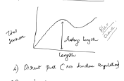
The transferred tendon should be set near an effective portion of its length–tension curve. Excessive tension restricts motion, while insufficient tension produces a weak transfer.
Principles of tendon transfer
- The donor muscle should be expendable
- Its strength should be adequate for the new function
- Joints must be supple before transfer
- The line of pull should be as direct as possible
- Excursion of the donor should match the required movement
- Avoid using a tendon that must perform several conflicting functions