Overview
Lateral condyle fractures are common paediatric elbow fractures and have a relatively high risk of displacement, nonunion and growth disturbance because the fracture extends into the joint and the fragment is exposed to deforming forces from the extensor-supinator origin.
Mechanism
Two mechanisms are described:
- Pull-off mechanism from traction of the common extensor origin
- Push-off mechanism from the radial head impacting the lateral condyle
Imaging
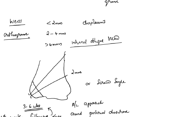
Obtain AP, lateral and internal oblique radiographs. The internal oblique view may demonstrate displacement not obvious on the AP view.
Classification by displacement
A practical treatment-based approach is:
- < 2 mm displacement, intact articular hinge: usually treated in a long-arm cast with close radiographic follow-up
- 2–4 mm displacement with preserved articular alignment: often suitable for closed reduction and percutaneous fixation
- > 4 mm displacement, rotation or articular incongruity: usually requires open reduction and internal fixation
Treatment principles
Non-operative
- Long-arm cast
- Close early radiographic surveillance because displacement may occur after presentation
Operative
- Percutaneous K-wire or screw fixation after satisfactory closed reduction
- Open reduction when the articular surface cannot be reduced reliably by closed means
Complications
- Delayed union or nonunion
- Cubitus valgus or varus
- Lateral condylar overgrowth
- Fishtail deformity
- Avascular necrosis
- Tardy ulnar nerve palsy in longstanding deformity
References
- Orthobullets. Lateral Condyle Fracture – Pediatric.