Gaucher disease
Gaucher disease is an autosomal recessive lysosomal storage disorder caused by deficiency of β-glucocerebrosidase, resulting in accumulation of glucocerebroside within macrophages.
Clinical features
Systemic manifestations include:
- Hepatosplenomegaly
- Anaemia
- Thrombocytopenia and easy bruising
- Growth disturbance in affected children
Orthopaedic manifestations include:
- Bone pain and acute bone crises
- Osteopenia
- Pathological fracture
- Medullary expansion and deformity
- Osteonecrosis, particularly of the femoral head
- Vertebral collapse in severe disease
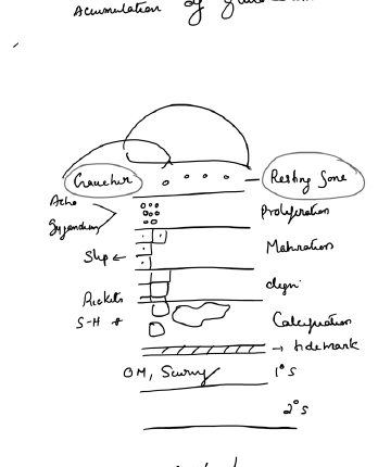
Radiographic features
The classic skeletal sign is the Erlenmeyer-flask deformity of the distal femur, caused by failure of normal metaphyseal remodelling. Other findings include osteopenia, medullary expansion, infarct-like change and vertebral involvement.
Diagnosis
Diagnosis is confirmed by reduced glucocerebrosidase enzyme activity, with molecular testing of GBA1 used for confirmation and counselling.
Treatment
- Enzyme replacement therapy for appropriate phenotypes
- Substrate-reduction therapy in selected patients
- Analgesia and treatment of bone crises
- Standard fracture care with attention to osteopenia and bleeding risk
- Arthroplasty or other reconstruction for advanced joint destruction or osteonecrosis when indicated
Duchenne muscular dystrophy
Duchenne muscular dystrophy is an X-linked recessive dystrophinopathy caused by mutations in the dystrophin gene that lead to absent or near-absent functional dystrophin.
Presentation
Symptoms usually begin in early childhood and include:
- Delayed motor milestones
- Progressive proximal muscle weakness
- Waddling or Trendelenburg gait
- Difficulty climbing stairs or rising from the floor
- Gowers sign
- Calf pseudohypertrophy
- Progressive loss of ambulation
Serum creatine kinase is markedly elevated. Diagnosis is established primarily by genetic testing; muscle biopsy is now reserved for selected unresolved cases.
Orthopaedic manifestations
- Hip, knee and ankle contractures
- Equinus or equinovarus foot deformity
- Progressive scoliosis, particularly after loss of ambulation
- Osteopenia and increased fracture risk
Management
Management is multidisciplinary and includes:
- Regular stretching and physiotherapy
- Night splints or AFOs when useful for ankle position
- Glucocorticoid therapy as part of neuromuscular management
- Cardiac and respiratory surveillance
- Bone-health surveillance
- Monitoring of spinal deformity
- Selective soft-tissue surgery for troublesome contractures
- Spinal fusion for progressive scoliosis when indicated and physiologically appropriate
Becker muscular dystrophy
Becker muscular dystrophy is also dystrophin-related but usually has an in-frame mutation with reduced or abnormal dystrophin. The phenotype is milder, with later onset and slower progression than Duchenne muscular dystrophy.