Compartments
The thigh is divided by intermuscular septa into anterior, medial and posterior compartments.
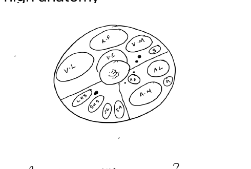
Anterior compartment
Main function: knee extension, with contribution to hip flexion.
Quadriceps femoris
- Rectus femoris: AIIS and supra-acetabular region to quadriceps tendon
- Vastus lateralis: greater trochanter and lateral linea aspera
- Vastus medialis: intertrochanteric line and medial linea aspera
- Vastus intermedius: anterior and lateral femoral shaft
All insert through the quadriceps tendon and patella into the tibial tubercle through the patellar tendon.
Nerve: femoral nerve.
Sartorius
- Origin: ASIS
- Insertion: pes anserinus on proximal medial tibia
- Nerve: femoral nerve
Medial compartment
Main function: hip adduction.
- Adductor longus: pubis to middle third of linea aspera
- Adductor brevis: pubis to proximal linea aspera
- Adductor magnus: pubic/ischial origin to linea aspera and adductor tubercle
- Gracilis: pubis to pes anserinus
- Obturator externus: obturator membrane to trochanteric fossa
Nerve: predominantly obturator nerve. The hamstring part of adductor magnus is supplied by the tibial division of the sciatic nerve.
Posterior compartment
Main functions: hip extension and knee flexion.
- Semitendinosus: ischial tuberosity to pes anserinus
- Semimembranosus: ischial tuberosity to posteromedial tibia
- Biceps femoris long head: ischial tuberosity to fibular head
- Biceps femoris short head: linea aspera to fibular head
Nerve: tibial division of sciatic nerve, except short head of biceps femoris, supplied by the common fibular division.
Femoral triangle
Boundaries:
- Superior: inguinal ligament
- Lateral: sartorius
- Medial: adductor longus
Contents from lateral to medial are femoral nerve, artery, vein and lymphatics.
Adductor canal
Contains the femoral artery and vein and the saphenous nerve. It runs from the apex of the femoral triangle to the adductor hiatus.
References
- Orthobullets. Thigh Anatomy.