Principles
Wrist arthroscopy portals are named according to their relationship to the extensor compartments. Safe portal placement requires accurate identification of the dorsal tendons and protection of superficial sensory nerves and vessels.
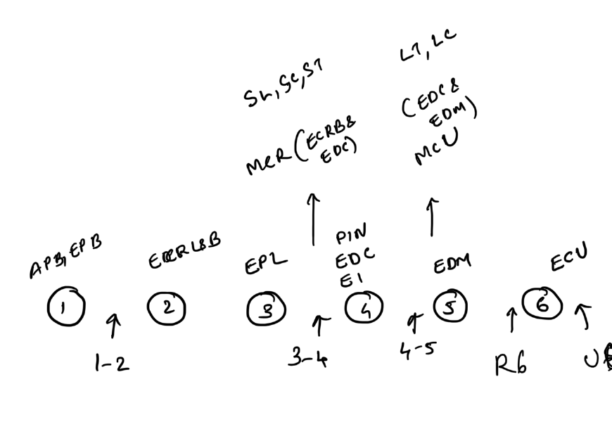
Radiocarpal portals
3–4 portal
- Between the EPL and EDC tendons
- Approximately 1 cm distal to Lister tubercle
- Usually the primary viewing portal
4–5 portal
- Between EDC and EDM
- Useful for instrumentation and TFCC visualisation
6R portal
- Just radial to the ECU tendon
- Useful for ulnar-sided visualisation and TFCC procedures
- Dorsal sensory branch of the ulnar nerve is at risk
6U portal
- Just ulnar to the ECU tendon
- Used selectively for ulnar-sided procedures
- Dorsal sensory branch of the ulnar nerve is at risk
1–2 portal
- Between the first and second extensor compartments in the anatomical snuffbox region
- Radial artery and superficial radial nerve are at risk
Midcarpal portals
Midcarpal radial
- Distal to the 3–4 portal
- Used to inspect the scapholunate, scaphocapitate and STT region
Midcarpal ulnar
- Distal to the 4–5 portal
- Used to inspect the lunotriquetral and ulnar midcarpal joints
First CMC portals
1R and 1U portals lie on either side of the EPB region at the thumb CMC joint and can be used for diagnostic or therapeutic basal-joint arthroscopy.
Safety points
- Mark all tendons and bony landmarks before traction.
- Make a small skin incision and bluntly dissect to capsule.
- Avoid blind deep sharp dissection.
- Protect the superficial radial nerve and dorsal sensory branch of the ulnar nerve.
References
- Orthobullets — Wrist Arthroscopy