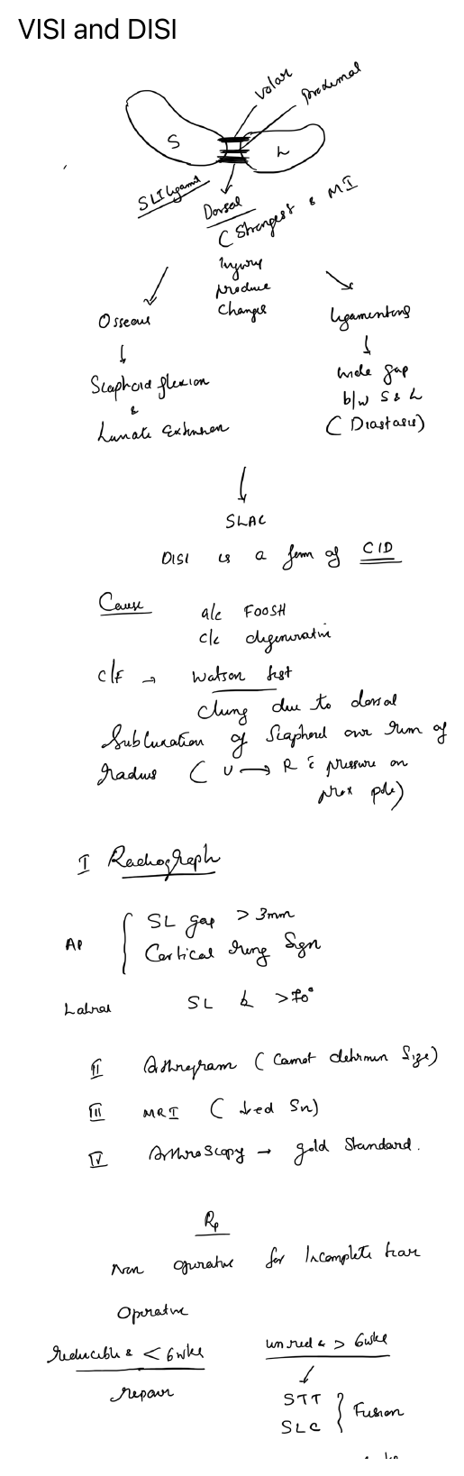
Definitions
These are patterns of carpal instability identified mainly on the lateral wrist radiograph.
- DISI — dorsal intercalated segment instability
- VISI — volar intercalated segment instability
The “intercalated segment” refers primarily to the lunate.
DISI
In DISI, the lunate extends dorsally.
Common cause
- Scapholunate ligament injury
- May follow trauma such as a fall on the outstretched hand
- May also occur with degenerative scapholunate dissociation
Associated mechanics
- Scaphoid tends to flex
- Lunate tends to extend dorsally
Radiographic features
- Scapholunate gap may be widened on AP view
- “Cortical ring sign” may be seen on the scaphoid
- Scapholunate angle > 70° on lateral view suggests DISI
VISI
In VISI, the lunate flexes volarly.
Common cause
- Lunotriquetral ligament injury
Radiographic features
- Scapholunate angle < 30° may be seen
- Lunate is tilted volarly on the lateral view
Investigations
- Plain radiographs
- Stress views in selected cases
- MRI for ligament assessment in selected cases
- Arthroscopy is the most accurate method for defining the ligament injury
Clinical significance
Untreated carpal instability may progress to chronic pain, weakness and degenerative change such as SLAC wrist in longstanding scapholunate instability.
Treatment principles
- Acute reducible injuries may be treated with repair and temporary stabilisation
- Chronic or irreducible cases may require reconstruction or salvage procedures
- Treatment depends on chronicity, reducibility and arthritic change
Diagram