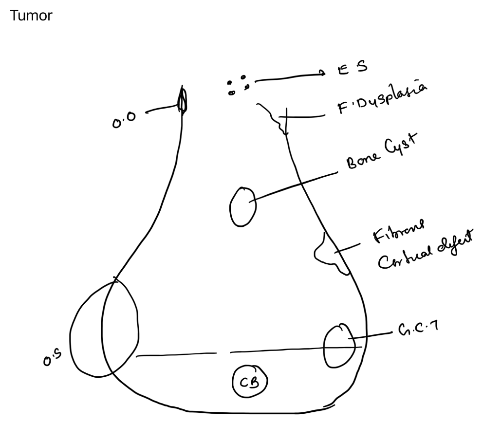
Importance of age and location
The differential diagnosis of a bone lesion depends strongly on:
- patient age
- bone involved
- epiphyseal, metaphyseal or diaphyseal location
- central versus eccentric position
- cortical versus medullary origin
Typical patterns
Epiphysis
- chondroblastoma in younger patients
- giant cell tumour after physeal closure
Metaphysis
- osteosarcoma
- simple bone cyst
- non-ossifying fibroma
- osteochondroma arises from the metaphyseal region and grows away from the joint
Diaphysis
- Ewing sarcoma
- fibrous dysplasia may involve the diaphysis or metaphysis
Cortical lesions
- osteoid osteoma
- non-ossifying fibroma / fibrous cortical defect
Radiographic approach
When interpreting a lesion, assess:
- margin and zone of transition
- periosteal reaction
- matrix mineralisation
- cortical destruction
- soft-tissue mass
- relationship to the physis and joint
Age and location narrow the differential, but aggressive imaging features and clinical context determine urgency.
Diagram